 Movie
Movie Controller

[English] 日本語
 Yorodumi
- PDB-4d9b: Pyridoxamine 5' phosphate (PMP) bound form of Salmonella typhimur... -
+ Open data

- Basic information
| Entry | Database: PDB / ID: 4d9b | ||||||
|---|---|---|---|---|---|---|---|
| Title | Pyridoxamine 5' phosphate (PMP) bound form of Salmonella typhimurium D-Cysteine desulfhydrase obtained after co-crystallization with D-cycloserine | ||||||
 Components Components | D-cysteine desulfhydrase | ||||||
 Keywords Keywords | LYASE / Fold type II PLP-dependent enzyme or tryptophan synthase beta subunit like family / PLP dependent enzyme | ||||||
| Function / homology |  Function and homology information Function and homology informationD-cysteine desulfhydrase / D-amino acid metabolic process / D-cysteine desulfhydrase activity Similarity search - Function | ||||||
| Biological species |  Salmonella typhimurium (bacteria) Salmonella typhimurium (bacteria) | ||||||
| Method |  X-RAY DIFFRACTION / SYNCHROTRON / MOLECULAR REPLACEMENT / Resolution: 1.67 Å X-RAY DIFFRACTION / SYNCHROTRON / MOLECULAR REPLACEMENT / Resolution: 1.67 Å | ||||||
 Authors Authors | Bharath, S.R. / Shveta, B. / Rajesh, K.H. / Savithri, H.S. / Murthy, M.R.N. | ||||||
 Citation Citation | Journal: Plos One / Year: 2012 Title: Structural and Mutational Studies on Substrate Specificity and Catalysis of Salmonella typhimurium D-Cysteine Desulfhydrase. Authors: Bharath, S.R. / Bisht, S. / Harijan, R.K. / Savithri, H.S. / Murthy, M.R.N. | ||||||
| History |
|
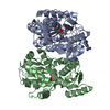
- Structure visualization
| Structure viewer | Molecule: MolmilJmol/JSmol |
|---|
- Downloads & links
-Download
| PDBx/mmCIF format | 4d9b.cif.gz | 271.7 KB | Display | PDBx/mmCIF format |
|---|---|---|---|---|
| PDB format | pdb4d9b.ent.gz | 218.2 KB | Display | PDB format |
| PDBx/mmJSON format | 4d9b.json.gz | Tree view | PDBx/mmJSON format | |
| Others |  Other downloads Other downloads |
-Validation report
| Arichive directory | https://data.pdbj.org/pub/pdb/validation_reports/d9/4d9bftp://data.pdbj.org/pub/pdb/validation_reports/d9/4d9b | HTTPS FTP |
|---|
-Related structure data
| Related structure data | 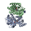 4d8tSC 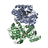 4d8uC  4d8wC  4d92C  4d96C  4d97C  4d99C  4d9cC  4d9eC  4d9fC S: Starting model for refinement C: citing same article ( |
|---|---|
| Similar structure data |


-Links
 PDBj
PDBj

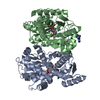
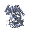
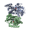
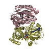
- Assembly
| Deposited unit | 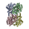
| ||||||||
|---|---|---|---|---|---|---|---|---|---|
| 1 | 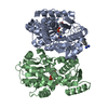
| ||||||||
| 2 | 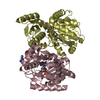
| ||||||||
| Unit cell |
|
-Components
| #1: Protein | Mass: 36550.727 Da / Num. of mol.: 4 Source method: isolated from a genetically manipulated source Source: (gene. exp.) Salmonella typhimurium (bacteria) / Gene: dcyD, STM1953 / Plasmid: pRSET C / Production host: #2: Chemical |   Mass: 120.152 Da / Num. of mol.: 3 / Source method: obtained synthetically / Formula: C7H8N2 Mass: 120.152 Da / Num. of mol.: 3 / Source method: obtained synthetically / Formula: C7H8N2#3: Chemical | ChemComp-PMP /   Mass: 248.173 Da / Num. of mol.: 4 / Source method: obtained synthetically / Formula: C8H13N2O5P Mass: 248.173 Da / Num. of mol.: 4 / Source method: obtained synthetically / Formula: C8H13N2O5P#4: Chemical | ChemComp-EDO /   Mass: 62.068 Da / Num. of mol.: 6 / Source method: obtained synthetically / Formula: C2H6O2 Mass: 62.068 Da / Num. of mol.: 6 / Source method: obtained synthetically / Formula: C2H6O2#5: Water | ChemComp-HOH / |  Mass: 18.015 Da / Num. of mol.: 887 / Source method: isolated from a natural source / Formula: H2O Mass: 18.015 Da / Num. of mol.: 887 / Source method: isolated from a natural source / Formula: H2O |
|---|
-Experimental details
-Experiment
| Experiment | Method: X-RAY DIFFRACTION / Number of used crystals: 1 |
|---|
- Sample preparation
| Crystal | Density Matthews: 2.24 Å3/Da / Density % sol: 44.98 % |
|---|---|
| Crystal grow | Temperature: 298 K / Method: vapor diffusion, hanging drop / pH: 8.6 Details: 1.5M Ammonium sulphate, 15% (v/v) ethylene glycol, 0.1M HEPES, 0.2% Benzamidine, 10mM D-Cycloserine, pH 8.6, VAPOR DIFFUSION, HANGING DROP, temperature 298K |
-Data collection
| Diffraction | Mean temperature: 100 K |
|---|---|
| Diffraction source | Source: SYNCHROTRON / Site: ESRF  / Beamline: BM14 / Beamline: BM14 |
| Detector | Type: MARMOSAIC 225 mm CCD / Detector: CCD / Date: Apr 19, 2010 / Details: bent collimating mirror and torroid |
| Radiation | Monochromator: Si(III) monochromator / Protocol: SINGLE WAVELENGTH / Monochromatic (M) / Laue (L): M / Scattering type: x-ray |
| Radiation wavelength | Relative weight: 1 |
| Reflection | Resolution: 1.67→50 Å / Num. obs: 143652 / % possible obs: 96.9 % / Redundancy: 4 % / Rmerge(I) obs: 0.053 / Net I/σ(I): 33.94 |
| Reflection shell | Resolution: 1.67→1.73 Å / Rmerge(I) obs: 0.271 / Mean I/σ(I) obs: 3.68 / % possible all: 89.9 |
- Processing
| Software |
| |||||||||||||||||||||||||||||||||||||||||||||||||||||||||||||||||
|---|---|---|---|---|---|---|---|---|---|---|---|---|---|---|---|---|---|---|---|---|---|---|---|---|---|---|---|---|---|---|---|---|---|---|---|---|---|---|---|---|---|---|---|---|---|---|---|---|---|---|---|---|---|---|---|---|---|---|---|---|---|---|---|---|---|---|
| Refinement | Method to determine structure: MOLECULAR REPLACEMENT Starting model: PDB ENTRY 4D8T Resolution: 1.67→39.91 Å / Cor.coef. Fo:Fc: 0.967 / Cor.coef. Fo:Fc free: 0.954 / SU B: 2.037 / SU ML: 0.068 / Cross valid method: THROUGHOUT / ESU R: 0.1 / ESU R Free: 0.099 / Stereochemistry target values: MAXIMUM LIKELIHOOD
| |||||||||||||||||||||||||||||||||||||||||||||||||||||||||||||||||
| Solvent computation | Ion probe radii: 0.8 Å / Shrinkage radii: 0.8 Å / VDW probe radii: 1.4 Å / Solvent model: BABINET MODEL WITH MASK | |||||||||||||||||||||||||||||||||||||||||||||||||||||||||||||||||
| Displacement parameters | Biso mean: 22.025 Å2
| |||||||||||||||||||||||||||||||||||||||||||||||||||||||||||||||||
| Refinement step | Cycle: LAST / Resolution: 1.67→39.91 Å
| |||||||||||||||||||||||||||||||||||||||||||||||||||||||||||||||||
| Refine LS restraints |
| |||||||||||||||||||||||||||||||||||||||||||||||||||||||||||||||||
| LS refinement shell | Resolution: 1.673→1.717 Å / Total num. of bins used: 20
|
