 Movie
Movie Controller

[English] 日本語
 Yorodumi
- PDB-4iz0: Crystal structure of HCV NS5B polymerase in complex with 2,4,5-tr... -
+ Open data

- Basic information
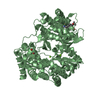
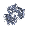
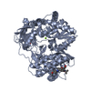
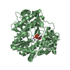
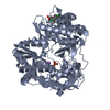
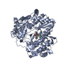
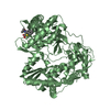
| Entry | Database: PDB / ID: 4iz0 | ||||||
|---|---|---|---|---|---|---|---|
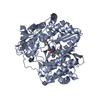
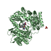
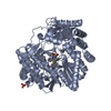
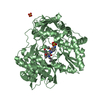
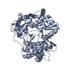
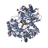
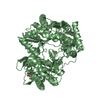
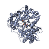
| Title | Crystal structure of HCV NS5B polymerase in complex with 2,4,5-trichloro-N-(5-methyl-1,2-oxazol-3-yl)benzenesulfonamide | ||||||
 Components Components | RNA-directed RNA polymerase | ||||||
 Keywords Keywords | TRANSFERASE/TRANSFERASE INHIBITOR / HCV NS5B / TRANSFERASE-TRANSFERASE INHIBITOR complex | ||||||
| Function / homology |  Function and homology information Function and homology informationhepacivirin / host cell mitochondrial membrane / host cell lipid droplet / symbiont-mediated transformation of host cell / symbiont-mediated suppression of host TRAF-mediated signal transduction / symbiont-mediated perturbation of host cell cycle G1/S transition checkpoint / symbiont-mediated suppression of host JAK-STAT cascade via inhibition of STAT1 activity / symbiont-mediated suppression of host cytoplasmic pattern recognition receptor signaling pathway via inhibition of MAVS activity / SH3 domain binding / nucleoside-triphosphate phosphatase ...hepacivirin / host cell mitochondrial membrane / host cell lipid droplet / symbiont-mediated transformation of host cell / symbiont-mediated suppression of host TRAF-mediated signal transduction / symbiont-mediated perturbation of host cell cycle G1/S transition checkpoint / symbiont-mediated suppression of host JAK-STAT cascade via inhibition of STAT1 activity / symbiont-mediated suppression of host cytoplasmic pattern recognition receptor signaling pathway via inhibition of MAVS activity / SH3 domain binding / nucleoside-triphosphate phosphatase / viral nucleocapsid / channel activity / monoatomic ion transmembrane transport / clathrin-dependent endocytosis of virus by host cell / molecular adaptor activity / Hydrolases; Acting on peptide bonds (peptidases); Cysteine endopeptidases / RNA helicase activity / host cell perinuclear region of cytoplasm / host cell endoplasmic reticulum membrane / RNA helicase / symbiont-mediated suppression of host type I interferon-mediated signaling pathway / ribonucleoprotein complex / serine-type endopeptidase activity / symbiont-mediated activation of host autophagy / RNA-directed RNA polymerase / cysteine-type endopeptidase activity / viral RNA genome replication / RNA-directed RNA polymerase activity / fusion of virus membrane with host endosome membrane / viral envelope / virion attachment to host cell / host cell nucleus / host cell plasma membrane / virion membrane / structural molecule activity / ATP hydrolysis activity / DNA-templated transcription / proteolysis / RNA binding / zinc ion binding / ATP binding Similarity search - Function | ||||||
| Biological species |  Hepatitis C virus Hepatitis C virus | ||||||
| Method |  X-RAY DIFFRACTION / MOLECULAR REPLACEMENT / Resolution: 2.22 Å X-RAY DIFFRACTION / MOLECULAR REPLACEMENT / Resolution: 2.22 Å | ||||||
 Authors Authors | Coulombe, R. | ||||||
 Citation Citation | Journal: Bioorg.Med.Chem.Lett. / Year: 2013 Title: Discovery of a novel series of non-nucleoside thumb pocket 2 HCV NS5B polymerase inhibitors. Authors: Stammers, T.A. / Coulombe, R. / Rancourt, J. / Thavonekham, B. / Fazal, G. / Goulet, S. / Jakalian, A. / Wernic, D. / Tsantrizos, Y. / Poupart, M.A. / Bos, M. / McKercher, G. / Thauvette, L. ...Authors: Stammers, T.A. / Coulombe, R. / Rancourt, J. / Thavonekham, B. / Fazal, G. / Goulet, S. / Jakalian, A. / Wernic, D. / Tsantrizos, Y. / Poupart, M.A. / Bos, M. / McKercher, G. / Thauvette, L. / Kukolj, G. / Beaulieu, P.L. | ||||||
| History |
|
- Structure visualization
| Structure viewer | Molecule: MolmilJmol/JSmol |
|---|
- Downloads & links
-Download
| PDBx/mmCIF format | 4iz0.cif.gz | 219.8 KB | Display | PDBx/mmCIF format |
|---|---|---|---|---|
| PDB format | pdb4iz0.ent.gz | 177 KB | Display | PDB format |
| PDBx/mmJSON format | 4iz0.json.gz | Tree view | PDBx/mmJSON format | |
| Others |  Other downloads Other downloads |
-Validation report
| Arichive directory | https://data.pdbj.org/pub/pdb/validation_reports/iz/4iz0ftp://data.pdbj.org/pub/pdb/validation_reports/iz/4iz0 | HTTPS FTP |
|---|
-Related structure data
| Related structure data |  4j02C 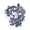 4j04C  4j06C  4j08C  4j0aC  3mwvS S: Starting model for refinement C: citing same article ( |
|---|---|
| Similar structure data |


-Links
 PDBj
PDBj
- Assembly
| Deposited unit | 
| ||||||||
|---|---|---|---|---|---|---|---|---|---|
| 1 | 
| ||||||||
| 2 | 
| ||||||||
| Unit cell |
|
-Components
| #1: Protein | Mass: 64285.637 Da / Num. of mol.: 2 / Fragment: UNP residues 2420-2989 Source method: isolated from a genetically manipulated source Source: (gene. exp.) Hepatitis C virus / Strain: HC-J4 / Gene: NS5B / Plasmid: pET29B / Production host:  #2: Chemical |   Mass: 341.598 Da / Num. of mol.: 2 / Source method: obtained synthetically / Formula: C10H7Cl3N2O3S Mass: 341.598 Da / Num. of mol.: 2 / Source method: obtained synthetically / Formula: C10H7Cl3N2O3S#3: Water | ChemComp-HOH / |  Mass: 18.015 Da / Num. of mol.: 4 / Source method: isolated from a natural source / Formula: H2O Mass: 18.015 Da / Num. of mol.: 4 / Source method: isolated from a natural source / Formula: H2OHas protein modification | Y | |
|---|
-Experimental details
-Experiment
| Experiment | Method: X-RAY DIFFRACTION / Number of used crystals: 1 |
|---|
- Sample preparation
| Crystal | Density Matthews: 2.97 Å3/Da / Density % sol: 58.58 % |
|---|---|
| Crystal grow | Temperature: 284 K / Method: vapor diffusion, hanging drop / pH: 5.4 Details: 100 mM MES, 21% PEG5000 MME, 200 mM ammonium sulfate, 10% glycerol, pH 5.4, VAPOR DIFFUSION, HANGING DROP, temperature 284K |
-Data collection
| Diffraction | Mean temperature: 110 K |
|---|---|
| Diffraction source | Source: ROTATING ANODE / Type: RIGAKU FR-E SUPERBRIGHT / Wavelength: 1.54 Å |
| Detector | Type: MAR scanner 345 mm plate / Detector: IMAGE PLATE / Date: Nov 29, 2004 / Details: hires2 |
| Radiation | Protocol: SINGLE WAVELENGTH / Monochromatic (M) / Laue (L): M / Scattering type: x-ray |
| Radiation wavelength | Wavelength: 1.54 Å / Relative weight: 1 |
| Reflection | Resolution: 2.22→40 Å / Num. obs: 75980 / % possible obs: 99.5 % / Observed criterion σ(I): 1 / Redundancy: 4.7 % / Rmerge(I) obs: 0.067 / Net I/σ(I): 19.65 |
| Reflection shell | Resolution: 2.22→2.3 Å / Redundancy: 3.9 % / Rmerge(I) obs: 0.477 / Mean I/σ(I) obs: 2.68 / Num. unique all: 7226 / % possible all: 95.9 |
- Processing
| Software |
| ||||||||||||||||||||
|---|---|---|---|---|---|---|---|---|---|---|---|---|---|---|---|---|---|---|---|---|---|
| Refinement | Method to determine structure: MOLECULAR REPLACEMENT Starting model: PDB ENTRY 3MWV Resolution: 2.22→40 Å / Occupancy max: 1 / Occupancy min: 1 / σ(F): 0
| ||||||||||||||||||||
| Displacement parameters | Biso max: 94.65 Å2 / Biso mean: 40.3372 Å2 / Biso min: 18.28 Å2
| ||||||||||||||||||||
| Refinement step | Cycle: LAST / Resolution: 2.22→40 Å
| ||||||||||||||||||||
| Refine LS restraints |
| ||||||||||||||||||||
| Xplor file |
|
