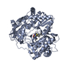
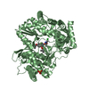
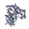
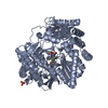
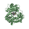
[English] 日本語
Yorodumi
- PDB-3upi: Synthesis of novel 4,5-dihydrofurano indoles and their evaluation... -

+
Open data


ID or keywords:

Loading...

-
Basic information

Entry
Database: PDB / ID: 3upi
TitleSynthesis of novel 4,5-dihydrofurano indoles and their evaluation as HCV NS5B polymerase inhibitors
ComponentsRNA-directed RNA polymeraseRNA-dependent RNA polymerase
KeywordsTRANSFERASE/TRANSFERASE INHIBITOR / TRANSFERASE-TRANSFERASE INHIBITOR complex
Function / homology
Function and homology information


hepacivirin / host cell mitochondrial membrane / host cell lipid droplet / symbiont-mediated suppression of host TRAF-mediated signal transduction / transformation of host cell by virus / symbiont-mediated perturbation of host cell cycle G1/S transition checkpoint / symbiont-mediated suppression of host JAK-STAT cascade via inhibition of STAT1 activity / symbiont-mediated suppression of host cytoplasmic pattern recognition receptor signaling pathway via inhibition of MAVS activity / SH3 domain binding / : ...hepacivirin / host cell mitochondrial membrane / host cell lipid droplet / symbiont-mediated suppression of host TRAF-mediated signal transduction / transformation of host cell by virus / symbiont-mediated perturbation of host cell cycle G1/S transition checkpoint / symbiont-mediated suppression of host JAK-STAT cascade via inhibition of STAT1 activity / symbiont-mediated suppression of host cytoplasmic pattern recognition receptor signaling pathway via inhibition of MAVS activity / SH3 domain binding / : / nucleoside-triphosphate phosphatase / protein complex oligomerization / monoatomic ion channel activity / clathrin-dependent endocytosis of virus by host cell / viral nucleocapsid / Hydrolases; Acting on peptide bonds (peptidases); Cysteine endopeptidases / RNA helicase activity / host cell endoplasmic reticulum membrane / host cell perinuclear region of cytoplasm / molecular adaptor activity / RNA helicase / induction by virus of host autophagy / RNA-directed RNA polymerase / ribonucleoprotein complex / viral RNA genome replication / cysteine-type endopeptidase activity / RNA-dependent RNA polymerase activity / serine-type endopeptidase activity / fusion of virus membrane with host endosome membrane / viral envelope / symbiont-mediated suppression of host type I interferon-mediated signaling pathway / host cell nucleus / structural molecule activity / virion attachment to host cell / host cell plasma membrane / virion membrane / ATP hydrolysis activity / proteolysis / RNA binding / zinc ion binding / ATP binding / membrane
Similarity search - Function
Hepatitis C virus core protein, chain A superfamily / Hepatitus C virus, Non-structural 5a protein, C-terminal / Hepatitis C virus NS5A, 1B domain superfamily / Hepatitis C virus non-structural protein NS2, C-terminal domain / Hepatitis C virus non-structural protein NS2, N-terminal domain / Hepatitis C virus non-structural protein NS2 / HCV NS5a protein C-terminal region / Hepatitis C virus, Non-structural protein NS4b / Hepatitis C virus, Core protein, N-terminal / Hepatitis C virus non-structural protein NS4b ...Hepatitis C virus core protein, chain A superfamily / Hepatitus C virus, Non-structural 5a protein, C-terminal / Hepatitis C virus NS5A, 1B domain superfamily / Hepatitis C virus non-structural protein NS2, C-terminal domain / Hepatitis C virus non-structural protein NS2, N-terminal domain / Hepatitis C virus non-structural protein NS2 / HCV NS5a protein C-terminal region / Hepatitis C virus, Non-structural protein NS4b / Hepatitis C virus, Core protein, N-terminal / Hepatitis C virus non-structural protein NS4b / Hepatitis C virus capsid protein / Hepatitis C virus, Non-structural protein NS2 / Hepatitis C virus, Non-structural 5a protein / Hepatitis C virus, Non-structural 5a protein, domain 1a / Hepatitis C virus non-structural 5a, 1B domain / NS5A domain 1a superfamily / Hepatitis C virus non-structural 5a protein membrane anchor / Hepatitis C virus non-structural 5a zinc finger domain / Hepatitis C virus non-structural 5a domain 1b / Hepacivirus nonstructural protein 2 (NS2) protease domain profile. / Hepatitis C virus, Non-structural protein NS4a / Hepatitis C virus non-structural protein NS4a / Hepatitis C virus, Core protein, C-terminal / Hepatitis C virus core protein / Hepatitis C virus, Non-structural protein E2/NS1 / Hepatitis C virus non-structural protein E2/NS1 / Hepatitis C virus, Envelope glycoprotein E1 / Hepatitis C virus envelope glycoprotein E1 / RNA dependent RNA polymerase, hepatitis C virus / Viral RNA dependent RNA polymerase / Hepatitis C virus, NS3 protease, Peptidase S29 / Hepatitis C virus NS3 protease / Hepacivirus/Pegivirus NS3 protease domain profile. / DEAD box, Flavivirus / Flavivirus DEAD domain / Superfamilies 1 and 2 helicase C-terminal domain profile. / Superfamilies 1 and 2 helicase ATP-binding type-1 domain profile. / DEAD-like helicases superfamily / Helicase, C-terminal / Helicase superfamily 1/2, ATP-binding domain / Reverse transcriptase/Diguanylate cyclase domain / RNA-directed RNA polymerase, catalytic domain / RdRp of positive ssRNA viruses catalytic domain profile. / Peptidase S1, PA clan, chymotrypsin-like fold / DNA/RNA polymerase superfamily / Peptidase S1, PA clan / P-loop containing nucleoside triphosphate hydrolase
Similarity search - Domain/homology
Chem-0C2 / PHOSPHATE ION / Genome polyprotein
Similarity search - Component
Biological speciesHepatitis C virus
MethodX-RAY DIFFRACTION / FOURIER SYNTHESIS / Resolution: 2 Å
AuthorsVelazquez, F. / Venkataraman, S. / Lesburg, C.A. / Duca, J.S. / Rosenblum, S.B. / Kozlowski, J.A. / Njoroge, F.G.
CitationJournal: Org.Lett. / Year: 2012
Title: Synthesis of New 4,5-Dihydrofuranoindoles and Their Evaluation as HCV NS5B Polymerase Inhibitors.
Authors: Velazquez, F. / Venkatraman, S. / Lesburg, C.A. / Duca, J. / Rosenblum, S.B. / Kozlowski, J.A. / Njoroge, F.G.
History
DepositionNov 18, 2011Deposition site: RCSB / Processing site: RCSB
Revision 1.0Jan 25, 2012Provider: repository / Type: Initial release
Revision 1.1Feb 1, 2012Group: Database references

-
Structure visualization

Structure viewerMolecule:
MolmilJmol/JSmol

Downloads & links

-
Assembly

Deposited unit
A: RNA-directed RNA polymerase
B: RNA-directed RNA polymerase
hetero molecules


Theoretical massNumber of molelcules
Total (without water)131,7625
Polymers130,6402
Non-polymers1,1223
Water16,826934
1
A: RNA-directed RNA polymerase
hetero molecules


Theoretical massNumber of molelcules
Total (without water)65,9283
Polymers65,3201
Non-polymers6082
Water181
TypeNameSymmetry operationNumber
identity operation1_555x,y,z1
MethodPISA
2
B: RNA-directed RNA polymerase
hetero molecules


Theoretical massNumber of molelcules
Total (without water)65,8332
Polymers65,3201
Non-polymers5141
Water181
TypeNameSymmetry operationNumber
identity operation1_555x,y,z1
MethodPISA
Unit cell
Length a, b, c (Å)89.816, 106.538, 134.274
Angle α, β, γ (deg.)90.00, 90.00, 90.00
Int Tables number19
Space group name H-MP212121

-
Components

#1: Protein RNA-directed RNA polymerase / RNA-dependent RNA polymerase / NS5B


Mass: 65319.805 Da / Num. of mol.: 2 / Fragment: UNP residues 2420-2989
Source method: isolated from a genetically manipulated source
Source: (gene. exp.) Hepatitis C virus / Strain: HC-J4 / Production host: Escherichia coli (E. coli) / References: UniProt: O92972, RNA-directed RNA polymerase
#2: Chemical ChemComp-PO4 / PHOSPHATE ION / Phosphate


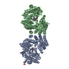
Mass: 94.971 Da / Num. of mol.: 1 / Source method: obtained synthetically / Formula: PO4
#3: Chemical ChemComp-0C2 / (3S)-6-(2,5-difluorobenzyl)-3-methyl-N-(methylsulfonyl)-8-(2-oxo-1,2-dihydropyridin-3-yl)-3,6-dihydro-2H-furo[2,3-e]indole-7-carboxamide


Mass: 513.513 Da / Num. of mol.: 2 / Source method: obtained synthetically / Formula: C25H21F2N3O5S
#4: Water ChemComp-HOH / water / Water


Mass: 18.015 Da / Num. of mol.: 934 / Source method: isolated from a natural source / Formula: H2O

-
Experimental details

-
Experiment

ExperimentMethod: X-RAY DIFFRACTION / Number of used crystals: 1

-
Sample preparation

CrystalDensity Matthews: 2.46 Å3/Da / Density % sol: 49.97 %
Crystal growTemperature: 300 K / Method: vapor diffusion, hanging drop / pH: 5.97
Details: 12.2% w/v PEG3350, 0.1 M sodium citrate, pH 5.97, VAPOR DIFFUSION, HANGING DROP, temperature 300K

-
Data collection

DiffractionMean temperature: 95 K
Diffraction sourceSource: ROTATING ANODE / Type: RIGAKU FR-E SUPERBRIGHT / Wavelength: 1.54 Å
DetectorType: RIGAKU RAXIS IV++ / Detector: IMAGE PLATE / Date: Feb 4, 2007
RadiationMonochromator: Varimax HR / Protocol: SINGLE WAVELENGTH / Monochromatic (M) / Laue (L): M / Scattering type: x-ray
Radiation wavelengthWavelength: 1.54 Å / Relative weight: 1
ReflectionResolution: 2→30 Å / Num. all: 87612 / Num. obs: 87525 / % possible obs: 99.9 % / Observed criterion σ(F): 1 / Observed criterion σ(I): 1 / Biso Wilson estimate: 37.91 Å2 / Rmerge(I) obs: 0.066 / Net I/σ(I): 8.9
Reflection shellResolution: 2→2.07 Å / Redundancy: 3.9 % / Rmerge(I) obs: 0.439 / Mean I/σ(I) obs: 2.2 / % possible all: 100

-
Processing

Software
NameVersionClassification
StructureStudiodata collection
BUSTER2.9.4refinement
d*TREKdata reduction
d*TREKdata scaling
BUSTER2.9.4phasing
RefinementMethod to determine structure: FOURIER SYNTHESIS / Resolution: 2→20 Å / Cor.coef. Fo:Fc: 0.9449 / Cor.coef. Fo:Fc free: 0.9228 / SU R Cruickshank DPI: 0.171 / Cross valid method: THROUGHOUT / σ(F): 0 / SU R Blow DPI: 0.182 / SU Rfree Blow DPI: 0.162 / SU Rfree Cruickshank DPI: 0.158
RfactorNum. reflection% reflectionSelection details
Rfree0.2355 4345 4.98 %RANDOM
Rwork0.1893 ---
all0.1916 87463 --
obs0.1916 87262 99.77 %-
Displacement parametersBiso mean: 41.36 Å2
Baniso -1Baniso -2Baniso -3
1--8.0814 Å20 Å20 Å2
2---0.6619 Å20 Å2
3---8.7433 Å2
Refine analyzeLuzzati coordinate error obs: 0.224 Å
Refinement stepCycle: LAST / Resolution: 2→20 Å
ProteinNucleic acidLigandSolventTotal
Num. atoms8735 0 77 934 9746
Refine LS restraints
Refine-IDTypeNumberRestraint functionWeightDev ideal
X-RAY DIFFRACTIONt_dihedral_angle_d3108SINUSOIDAL2
X-RAY DIFFRACTIONt_trig_c_planes222HARMONIC2
X-RAY DIFFRACTIONt_gen_planes1569HARMONIC5
X-RAY DIFFRACTIONt_it9129HARMONIC20
X-RAY DIFFRACTIONt_nbd
X-RAY DIFFRACTIONt_improper_torsion
X-RAY DIFFRACTIONt_pseud_angle
X-RAY DIFFRACTIONt_chiral_improper_torsion1206SEMIHARMONIC5
X-RAY DIFFRACTIONt_sum_occupancies
X-RAY DIFFRACTIONt_utility_distance
X-RAY DIFFRACTIONt_utility_angle
X-RAY DIFFRACTIONt_utility_torsion
X-RAY DIFFRACTIONt_ideal_dist_contact11317SEMIHARMONIC4
X-RAY DIFFRACTIONt_bond_d9129HARMONIC20.01
X-RAY DIFFRACTIONt_angle_deg12422HARMONIC21.02
X-RAY DIFFRACTIONt_omega_torsion3.14
X-RAY DIFFRACTIONt_other_torsion15.52
LS refinement shellResolution: 2→2.05 Å / Total num. of bins used: 20
RfactorNum. reflection% reflection
Rfree0.2612 307 4.81 %
Rwork0.2193 6076 -
all0.2214 6383 -
obs--99.77 %

+
About Yorodumi

-
News

-
Feb 9, 2022. New format data for meta-information of EMDB entries

New format data for meta-information of EMDB entries

  • Version 3 of the EMDB header file is now the official format.
  • The previous official version 1.9 will be removed from the archive.

Related info.:EMDB header

External links:wwPDB to switch to version 3 of the EMDB data model

-
Aug 12, 2020. Covid-19 info

Covid-19 info

URL: https://pdbj.org/emnavi/covid19.php

New page: Covid-19 featured information page in EM Navigator.

Related info.:Covid-19 info / Mar 5, 2020. Novel coronavirus structure data

+
Mar 5, 2020. Novel coronavirus structure data

Novel coronavirus structure data

Related info.:Yorodumi Speices / Aug 12, 2020. Covid-19 info

External links:COVID-19 featured content - PDBj / Molecule of the Month (242):Coronavirus Proteases

+
Jan 31, 2019. EMDB accession codes are about to change! (news from PDBe EMDB page)

EMDB accession codes are about to change! (news from PDBe EMDB page)

  • The allocation of 4 digits for EMDB accession codes will soon come to an end. Whilst these codes will remain in use, new EMDB accession codes will include an additional digit and will expand incrementally as the available range of codes is exhausted. The current 4-digit format prefixed with “EMD-” (i.e. EMD-XXXX) will advance to a 5-digit format (i.e. EMD-XXXXX), and so on. It is currently estimated that the 4-digit codes will be depleted around Spring 2019, at which point the 5-digit format will come into force.
  • The EM Navigator/Yorodumi systems omit the EMD- prefix.

Related info.:Q: What is EMD? / ID/Accession-code notation in Yorodumi/EM Navigator

External links:EMDB Accession Codes are Changing Soon! / Contact to PDBj

+
Jul 12, 2017. Major update of PDB

Major update of PDB

  • wwPDB released updated PDB data conforming to the new PDBx/mmCIF dictionary.
  • This is a major update changing the version number from 4 to 5, and with Remediation, in which all the entries are updated.
  • In this update, many items about electron microscopy experimental information are reorganized (e.g. em_software).
  • Now, EM Navigator and Yorodumi are based on the updated data.

External links:wwPDB Remediation / Enriched Model Files Conforming to OneDep Data Standards Now Available in the PDB FTP Archive

-
Yorodumi

Thousand views of thousand structures

  • Yorodumi is a browser for structure data from EMDB, PDB, SASBDB, etc.
  • This page is also the successor to EM Navigator detail page, and also detail information page/front-end page for Omokage search.
  • The word "yorodu" (or yorozu) is an old Japanese word meaning "ten thousand". "mi" (miru) is to see.

Related info.:EMDB / PDB / SASBDB / Comparison of 3 databanks / Yorodumi Search / Aug 31, 2016. New EM Navigator & Yorodumi / Yorodumi Papers / Jmol/JSmol / Function and homology information / Changes in new EM Navigator and Yorodumi

Read more