 Movie
Movie Controller

[English] 日本語
 Yorodumi
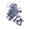
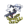
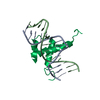
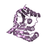
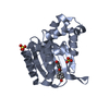
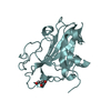
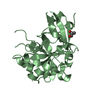
- PDB-3b90: Crystal Structure of the Catalytic Domain of Pectate Lyase PelI f... -
+ Open data

- Basic information
| Entry | Database: PDB / ID: 3b90 | ||||||
|---|---|---|---|---|---|---|---|
| Title | Crystal Structure of the Catalytic Domain of Pectate Lyase PelI from Erwinia chrysanthemi | ||||||
 Components Components | Endo-pectate lyase | ||||||
 Keywords Keywords | LYASE / Pectate Lyase / Pectin / Galacturonic Acid / Erwinia chrysanthemi / right-handed parallel beta helix fold / catalytic domain | ||||||
| Function / homology |  Function and homology information Function and homology informationpectate lyase / pectate lyase activity / pectin catabolic process / extracellular region / metal ion binding Similarity search - Function | ||||||
| Biological species |  Erwinia chrysanthemi (bacteria) Erwinia chrysanthemi (bacteria) | ||||||
| Method |  X-RAY DIFFRACTION / SYNCHROTRON / MOLECULAR REPLACEMENT / Resolution: 2.11 Å X-RAY DIFFRACTION / SYNCHROTRON / MOLECULAR REPLACEMENT / Resolution: 2.11 Å | ||||||
 Authors Authors | Creze, C. / Castang, S. / Derivery, E. / Haser, R. / Shevchik, V. / Gouet, P. | ||||||
 Citation Citation | Journal: J.Biol.Chem. / Year: 2008 Title: The Crystal Structure of Pectate Lyase PelI from Soft Rot Pathogen Erwinia chrysanthemi in Complex with Its Substrate Authors: Creze, C. / Castang, S. / Derivery, E. / Haser, R. / Hugouvieux-Cotte-Pattat, N. / Shevchik, V.E. / Gouet, P. | ||||||
| History |
|
- Structure visualization
| Structure viewer | Molecule: MolmilJmol/JSmol |
|---|
- Downloads & links
-Download
| PDBx/mmCIF format | 3b90.cif.gz | 105.1 KB | Display | PDBx/mmCIF format |
|---|---|---|---|---|
| PDB format | pdb3b90.ent.gz | 79.1 KB | Display | PDB format |
| PDBx/mmJSON format | 3b90.json.gz | Tree view | PDBx/mmJSON format | |
| Others |  Other downloads Other downloads |
-Validation report
| Arichive directory | https://data.pdbj.org/pub/pdb/validation_reports/b9/3b90ftp://data.pdbj.org/pub/pdb/validation_reports/b9/3b90 | HTTPS FTP |
|---|
-Related structure data


-Links
 PDBj
PDBj- Assembly
| Deposited unit | 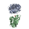
| ||||||||
|---|---|---|---|---|---|---|---|---|---|
| 1 | 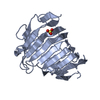
| ||||||||
| 2 | 
| ||||||||
| 3 | 
| ||||||||
| 4 | 
| ||||||||
| Unit cell |
|
-Components
| #1: Protein | Mass: 23851.688 Da / Num. of mol.: 2 Source method: isolated from a genetically manipulated source Source: (gene. exp.) Erwinia chrysanthemi (bacteria) / Genus: Dickeya / Gene: pelI / Plasmid: pT7-6 / Species (production host): Escherichia coli / Production host: #2: Chemical |   Mass: 40.078 Da / Num. of mol.: 2 / Source method: obtained synthetically / Formula: Ca Mass: 40.078 Da / Num. of mol.: 2 / Source method: obtained synthetically / Formula: Ca#3: Chemical | ChemComp-ZN /   Mass: 65.409 Da / Num. of mol.: 4 / Source method: obtained synthetically / Formula: Zn Mass: 65.409 Da / Num. of mol.: 4 / Source method: obtained synthetically / Formula: Zn#4: Chemical |   Mass: 96.063 Da / Num. of mol.: 2 / Source method: obtained synthetically / Formula: SO4 Mass: 96.063 Da / Num. of mol.: 2 / Source method: obtained synthetically / Formula: SO4#5: Water | ChemComp-HOH / |  Mass: 18.015 Da / Num. of mol.: 325 / Source method: isolated from a natural source / Formula: H2O Mass: 18.015 Da / Num. of mol.: 325 / Source method: isolated from a natural source / Formula: H2OHas protein modification | Y | Sequence details | THE AUTHORS STATE THAT AT THIS POSITION IT IS INDEED ARG171 AND THE DATABASE IS INCORRECT. | |
|---|
-Experimental details
-Experiment
| Experiment | Method: X-RAY DIFFRACTION / Number of used crystals: 1 |
|---|
- Sample preparation
| Crystal | Density Matthews: 1.91 Å3/Da / Density % sol: 35.53 % |
|---|---|
| Crystal grow | Temperature: 292 K / Method: vapor diffusion, hanging drop / pH: 6.5 Details: 0.01 M zinc sulfate heptahydrate, 0.1 M MES, 25% PEG 550, pH 6.5, VAPOR DIFFUSION, HANGING DROP, temperature 292K |
-Data collection
| Diffraction | Mean temperature: 100 K |
|---|---|
| Diffraction source | Source: SYNCHROTRON / Site: ESRF  / Beamline: ID14-3 / Wavelength: 0.931 Å / Beamline: ID14-3 / Wavelength: 0.931 Å |
| Detector | Type: MAR CCD 165 mm / Detector: CCD / Date: Nov 4, 2004 |
| Radiation | Protocol: SINGLE WAVELENGTH / Monochromatic (M) / Laue (L): M / Scattering type: x-ray |
| Radiation wavelength | Wavelength: 0.931 Å / Relative weight: 1 |
| Reflection | Resolution: 2.1→20 Å / Num. all: 21791 / Num. obs: 20021 / % possible obs: 91.9 % / Observed criterion σ(F): 0 / Observed criterion σ(I): -3 / Redundancy: 3.6 % / Biso Wilson estimate: 6.3 Å2 / Rmerge(I) obs: 0.108 / Rsym value: 0.108 / Net I/σ(I): 8.2 |
| Reflection shell | Resolution: 2.1→2.2 Å / Redundancy: 3.3 % / Rmerge(I) obs: 0.186 / Mean I/σ(I) obs: 5.3 / Num. unique all: 2172 / Rsym value: 0.186 / % possible all: 77.8 |
- Processing
| Software |
| ||||||||||||||||||||||||||||||||||||
|---|---|---|---|---|---|---|---|---|---|---|---|---|---|---|---|---|---|---|---|---|---|---|---|---|---|---|---|---|---|---|---|---|---|---|---|---|---|
| Refinement | Method to determine structure: MOLECULAR REPLACEMENT / Resolution: 2.11→19.83 Å / Rfactor Rfree error: 0.009 / Data cutoff high absF: 1891203.31 / Data cutoff low absF: 0 / Isotropic thermal model: RESTRAINED / Cross valid method: THROUGHOUT / σ(F): 0 / σ(I): -3 / Stereochemistry target values: Engh & Huber / Details: BULK SOLVENT MODEL USED
| ||||||||||||||||||||||||||||||||||||
| Solvent computation | Solvent model: FLAT MODEL / Bsol: 37.7373 Å2 / ksol: 0.35 e/Å3 | ||||||||||||||||||||||||||||||||||||
| Displacement parameters | Biso mean: 18.3 Å2
| ||||||||||||||||||||||||||||||||||||
| Refine analyze |
| ||||||||||||||||||||||||||||||||||||
| Refinement step | Cycle: LAST / Resolution: 2.11→19.83 Å
| ||||||||||||||||||||||||||||||||||||
| Refine LS restraints |
| ||||||||||||||||||||||||||||||||||||
| LS refinement shell | Resolution: 2.11→2.23 Å / Rfactor Rfree error: 0.025 / Total num. of bins used: 6
| ||||||||||||||||||||||||||||||||||||
| Xplor file |
|
