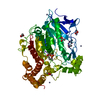
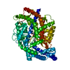
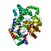
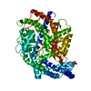
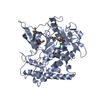
Hydrolases; Acting on peptide bonds (peptidases); Metalloendopeptidases / metallopeptidase activity / proteolysis / metal ion binding / plasma membrane Similarity search - Function
Uncharacterised conserved protein UCP006404, peptidase M50/CBS / Peptidase M50 / Peptidase family M50 / Domain in cystathionine beta-synthase and other proteins. / CBS domain superfamily / CBS domain / CBS domain / CBS domain profile. / Neutral zinc metallopeptidases, zinc-binding region signature. Similarity search - Domain/homology
Mass: 65.409 Da / Num. of mol.: 2 / Source method: obtained synthetically / Formula: Zn
-
Experimental details
-
Experiment
Experiment
Method: X-RAY DIFFRACTION / Number of used crystals: 2
-
Sample preparation
Crystal
Density Matthews: 4.08 Å3/Da / Density % sol: 69.87 %
Crystal grow
Temperature: 295 K / Method: vapor diffusion, hanging drop / pH: 8.8 Details: 0.1 M Tris (pH 8.8), 20% (w/v) PEG400, 50 mM NaCl, 5 mM CaCl2 and 30 mM MgCl2, VAPOR DIFFUSION, HANGING DROP, temperature 295K
-
Data collection
Diffraction
ID
Mean temperature (K)
Crystal-ID
1
100
1
2
100
1
Diffraction source
Source
Site
Beamline
ID
Wavelength (Å)
SYNCHROTRON
NSLS
X29A
1
1.0809
SYNCHROTRON
NSLS
X29A
2
0.97909, 0.97925
Detector
Type
ID
Detector
Date
ADSC QUANTUM 315
1
CCD
Aug 1, 2007
ADSC QUANTUM 315
2
CCD
Sep 17, 2007
Radiation
ID
Monochromator
Protocol
Monochromatic (M) / Laue (L)
Scattering type
Wavelength-ID
1
Si(111)
SINGLEWAVELENGTH
M
x-ray
1
2
Si(111)
MAD
M
x-ray
2
Radiation wavelength
ID
Wavelength (Å)
Relative weight
1
1.0809
1
2
0.97909
1
3
0.97925
1
Reflection
Resolution: 3.3→50 Å / Num. obs: 11853 / % possible obs: 99.3 % / Redundancy: 3.7 % / Biso Wilson estimate: 96.9 Å2 / Rsym value: 0.055 / Net I/σ(I): 14
Reflection shell
Resolution: 3.3→3.42 Å / Redundancy: 3.6 % / Mean I/σ(I) obs: 2.4 / Rsym value: 0.485 / % possible all: 99.8
-
Processing
Software
Name
Version
Classification
CNS
1.1
refinement
CBASS
datacollection
DENZO
datareduction
SCALEPACK
datascaling
SHELXS
phasing
Refinement
Method to determine structure: MAD / Resolution: 3.3→34.99 Å / Rfactor Rfree error: 0.013 / Data cutoff high absF: 1526734.75 / Data cutoff low absF: 0 / Isotropic thermal model: RESTRAINED / Cross valid method: THROUGHOUT / σ(F): 0 / Stereochemistry target values: Engh & Huber
In the structure databanks used in Yorodumi, some data are registered as the other names, "COVID-19 virus" and "2019-nCoV". Here are the details of the virus and the list of structure data.
Jan 31, 2019. EMDB accession codes are about to change! (news from PDBe EMDB page)
EMDB accession codes are about to change! (news from PDBe EMDB page)
The allocation of 4 digits for EMDB accession codes will soon come to an end. Whilst these codes will remain in use, new EMDB accession codes will include an additional digit and will expand incrementally as the available range of codes is exhausted. The current 4-digit format prefixed with “EMD-” (i.e. EMD-XXXX) will advance to a 5-digit format (i.e. EMD-XXXXX), and so on. It is currently estimated that the 4-digit codes will be depleted around Spring 2019, at which point the 5-digit format will come into force.
The EM Navigator/Yorodumi systems omit the EMD- prefix.
Related info.:Q: What is EMD? / ID/Accession-code notation in Yorodumi/EM Navigator
Yorodumi is a browser for structure data from EMDB, PDB, SASBDB, etc.
This page is also the successor to EM Navigator detail page, and also detail information page/front-end page for Omokage search.
The word "yorodu" (or yorozu) is an old Japanese word meaning "ten thousand". "mi" (miru) is to see.
Related info.:EMDB / PDB / SASBDB / Comparison of 3 databanks / Yorodumi Search / Aug 31, 2016. New EM Navigator & Yorodumi / Yorodumi Papers / Jmol/JSmol / Function and homology information / Changes in new EM Navigator and Yorodumi
 Movie
Movie Controller


 Open data
 Basic information
 Components
 Keywords
 Function and homology information
 Methanocaldococcus jannaschii (archaea)
 X-RAY DIFFRACTION /
  Authors
 Citation
 Structure visualization
 Downloads & links
 Other downloads
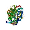

 PDBj
PDBj
 Assembly
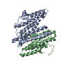


 Mass: 65.409 Da / Num. of mol.: 2 / Source method: obtained synthetically / Formula: Zn
 Sample preparation
 Processing