 Movie
Movie Controller

[English] 日本語
 Yorodumi
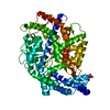
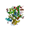
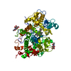
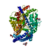
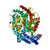
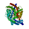
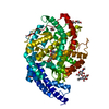
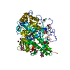
- PDB-6s1z: Crystal structure of Anopheles gambiae AnoACE2 in complex with fo... -
+ Open data

- Basic information
| Entry | Database: PDB / ID: 6s1z | |||||||||
|---|---|---|---|---|---|---|---|---|---|---|
| Title | Crystal structure of Anopheles gambiae AnoACE2 in complex with fosinoprilat | |||||||||
 Components Components | Angiotensin-converting enzyme | |||||||||
 Keywords Keywords | HYDROLASE / metalloprotease / mosquito control / insecticide design / fosinoprilat | |||||||||
| Function / homology |  Function and homology information Function and homology informationmetallodipeptidase activity / Hydrolases; Acting on peptide bonds (peptidases) / peptidyl-dipeptidase activity / carboxypeptidase activity / proteolysis / zinc ion binding / membrane Similarity search - Function | |||||||||
| Biological species |  | |||||||||
| Method |  X-RAY DIFFRACTION / SYNCHROTRON / MOLECULAR REPLACEMENT / Resolution: 2.5 Å X-RAY DIFFRACTION / SYNCHROTRON / MOLECULAR REPLACEMENT / Resolution: 2.5 Å | |||||||||
 Authors Authors | Cozier, G.E. / Acharya, K.R. / Cashman, J.S. | |||||||||
 Citation Citation | Journal: Biochem.J. / Year: 2019 Title: Crystal structures of angiotensin-converting enzyme from Anopheles gambiae in its native form and with a bound inhibitor. Authors: Cashman, J.S. / Cozier, G.E. / Harrison, C. / Isaac, R.E. / Acharya, K.R. | |||||||||
| History |
|
- Structure visualization
| Structure viewer | Molecule: MolmilJmol/JSmol |
|---|
- Downloads & links
-Download
| PDBx/mmCIF format | 6s1z.cif.gz | 240.1 KB | Display | PDBx/mmCIF format |
|---|---|---|---|---|
| PDB format | pdb6s1z.ent.gz | 193.4 KB | Display | PDB format |
| PDBx/mmJSON format | 6s1z.json.gz | Tree view | PDBx/mmJSON format | |
| Others |  Other downloads Other downloads |
-Validation report
| Arichive directory | https://data.pdbj.org/pub/pdb/validation_reports/s1/6s1zftp://data.pdbj.org/pub/pdb/validation_reports/s1/6s1z | HTTPS FTP |
|---|
-Related structure data
| Related structure data |  6s1ySC S: Starting model for refinement C: citing same article ( |
|---|---|
| Similar structure data |
-Links
 PDBj
PDBj

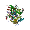
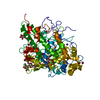
- Assembly
| Deposited unit | 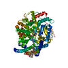
| ||||||||
|---|---|---|---|---|---|---|---|---|---|
| 1 |
| ||||||||
| Unit cell |
| ||||||||
| Components on special symmetry positions |
|
-Components
| #1: Protein | Mass: 72041.211 Da / Num. of mol.: 1 Source method: isolated from a genetically manipulated source Source: (gene. exp.) Gene: ANCE2, AgaP_AGAP009751 / Production host:  Spodoptera frugiperda (fall armyworm) Spodoptera frugiperda (fall armyworm)References: UniProt: A0NFU8, Hydrolases; Acting on peptide bonds (peptidases) |
|---|---|
| #2: Polysaccharide | beta-D-mannopyranose-(1-4)-2-acetamido-2-deoxy-beta-D-glucopyranose-(1-4)-2-acetamido-2-deoxy-beta- ...beta-D-mannopyranose-(1-4)-2-acetamido-2-deoxy-beta-D-glucopyranose-(1-4)-2-acetamido-2-deoxy-beta-D-glucopyranose Source method: isolated from a genetically manipulated source |
| #3: Chemical | ChemComp-KS8 / 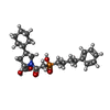  Mass: 435.494 Da / Num. of mol.: 1 / Source method: obtained synthetically / Formula: C23H34NO5P / Feature type: SUBJECT OF INVESTIGATION Mass: 435.494 Da / Num. of mol.: 1 / Source method: obtained synthetically / Formula: C23H34NO5P / Feature type: SUBJECT OF INVESTIGATION |
| #4: Chemical | ChemComp-ZN /   Mass: 65.409 Da / Num. of mol.: 1 / Source method: obtained synthetically / Formula: Zn Mass: 65.409 Da / Num. of mol.: 1 / Source method: obtained synthetically / Formula: Zn |
| #5: Water | ChemComp-HOH /  Mass: 18.015 Da / Num. of mol.: 25 / Source method: isolated from a natural source / Formula: H2O Mass: 18.015 Da / Num. of mol.: 25 / Source method: isolated from a natural source / Formula: H2O |
| Has ligand of interest | Y |
| Has protein modification | Y |
-Experimental details
-Experiment
| Experiment | Method: X-RAY DIFFRACTION / Number of used crystals: 1 |
|---|
- Sample preparation
| Crystal | Density Matthews: 3.27 Å3/Da / Density % sol: 62.39 % |
|---|---|
| Crystal grow | Temperature: 291 K / Method: vapor diffusion, sitting drop / pH: 7.8 Details: 43.2% v/v polypropylene glycol,30 mM sodium formate, 10 mM Tri pH 7.8, 0.3 % w/v gamma-PGA, 0.3% w/v PEG 20,000 |
-Data collection
| Diffraction | Mean temperature: 100 K / Serial crystal experiment: N |
|---|---|
| Diffraction source | Source: SYNCHROTRON / Site: Diamond  / Beamline: I04 / Wavelength: 0.9795 Å / Beamline: I04 / Wavelength: 0.9795 Å |
| Detector | Type: DECTRIS PILATUS3 6M / Detector: PIXEL / Date: Jan 19, 2018 |
| Radiation | Protocol: SINGLE WAVELENGTH / Monochromatic (M) / Laue (L): M / Scattering type: x-ray |
| Radiation wavelength | Wavelength: 0.9795 Å / Relative weight: 1 |
| Reflection | Resolution: 2.5→117.46 Å / Num. obs: 33809 / % possible obs: 100 % / Redundancy: 77.3 % / CC1/2: 1 / Rpim(I) all: 0.017 / Net I/σ(I): 24.8 |
| Reflection shell | Resolution: 2.5→2.6 Å / Redundancy: 79.8 % / Mean I/σ(I) obs: 4.7 / Num. unique obs: 3747 / CC1/2: 0.622 / Rpim(I) all: 0.137 / % possible all: 100 |
- Processing
| Software |
| |||||||||||||||||||||||||||||||||||||||||||||||||||||||||||||||||||||||||||||||||||||||||||
|---|---|---|---|---|---|---|---|---|---|---|---|---|---|---|---|---|---|---|---|---|---|---|---|---|---|---|---|---|---|---|---|---|---|---|---|---|---|---|---|---|---|---|---|---|---|---|---|---|---|---|---|---|---|---|---|---|---|---|---|---|---|---|---|---|---|---|---|---|---|---|---|---|---|---|---|---|---|---|---|---|---|---|---|---|---|---|---|---|---|---|---|---|
| Refinement | Method to determine structure: MOLECULAR REPLACEMENT Starting model: 6S1Y Resolution: 2.5→72.191 Å / SU ML: 0.51 / Cross valid method: FREE R-VALUE / σ(F): 1.76 / Phase error: 39.13
| |||||||||||||||||||||||||||||||||||||||||||||||||||||||||||||||||||||||||||||||||||||||||||
| Solvent computation | Shrinkage radii: 0.9 Å / VDW probe radii: 1.11 Å | |||||||||||||||||||||||||||||||||||||||||||||||||||||||||||||||||||||||||||||||||||||||||||
| Refinement step | Cycle: LAST / Resolution: 2.5→72.191 Å
| |||||||||||||||||||||||||||||||||||||||||||||||||||||||||||||||||||||||||||||||||||||||||||
| Refine LS restraints |
| |||||||||||||||||||||||||||||||||||||||||||||||||||||||||||||||||||||||||||||||||||||||||||
| LS refinement shell |
|
