 Movie
Movie Controller

[English] 日本語
 Yorodumi
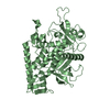
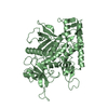
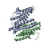
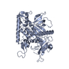
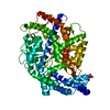
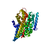
- PDB-6g16: Structure of the human RBBP4:MTA1(464-546) complex showing loop e... -
+ Open data

- Basic information
| Entry | Database: PDB / ID: 6g16 | ||||||
|---|---|---|---|---|---|---|---|
| Title | Structure of the human RBBP4:MTA1(464-546) complex showing loop exchange | ||||||
 Components Components |
| ||||||
 Keywords Keywords | TRANSCRIPTION / WD40 / corepressor / metastasis | ||||||
| Function / homology |  Function and homology information Function and homology informationCAF-1 complex / NURF complex / NuRD complex / regulation of cell fate specification / negative regulation of stem cell population maintenance / DNA replication-dependent chromatin assembly / ESC/E(Z) complex / Transcription of E2F targets under negative control by p107 (RBL1) and p130 (RBL2) in complex with HDAC1 / regulation of stem cell differentiation / Polo-like kinase mediated events ...CAF-1 complex / NURF complex / NuRD complex / regulation of cell fate specification / negative regulation of stem cell population maintenance / DNA replication-dependent chromatin assembly / ESC/E(Z) complex / Transcription of E2F targets under negative control by p107 (RBL1) and p130 (RBL2) in complex with HDAC1 / regulation of stem cell differentiation / Polo-like kinase mediated events / Transcription of E2F targets under negative control by DREAM complex / positive regulation of protein autoubiquitination / ATPase complex / entrainment of circadian clock by photoperiod / response to ionizing radiation / G1/S-Specific Transcription / locomotor rhythm / negative regulation of gene expression, epigenetic / Sin3-type complex / histone deacetylase complex / positive regulation of stem cell population maintenance / Transcriptional Regulation by E2F6 / SUMOylation of transcription factors / RNA Polymerase I Transcription Initiation / G0 and Early G1 / Cyclin E associated events during G1/S transition / Transcriptional regulation of brown and beige adipocyte differentiation by EBF2 / Cyclin A:Cdk2-associated events at S phase entry / Regulation of TP53 Activity through Acetylation / Deposition of new CENPA-containing nucleosomes at the centromere / negative regulation of cell migration / Regulation of PTEN gene transcription / ERCC6 (CSB) and EHMT2 (G9a) positively regulate rRNA expression / PRC2 methylates histones and DNA / Regulation of endogenous retroelements by KRAB-ZFP proteins / Defective pyroptosis / circadian regulation of gene expression / HDACs deacetylate histones / Regulation of endogenous retroelements by Piwi-interacting RNAs (piRNAs) / negative regulation of transforming growth factor beta receptor signaling pathway / brain development / Negative Regulation of CDH1 Gene Transcription / PKMTs methylate histone lysines / Activation of anterior HOX genes in hindbrain development during early embryogenesis / histone deacetylase binding / HCMV Early Events / transcription corepressor activity / nuclear envelope / double-strand break repair / nucleosome assembly / histone binding / Oxidative Stress Induced Senescence / Potential therapeutics for SARS / microtubule / RNA polymerase II-specific DNA-binding transcription factor binding / proteasome-mediated ubiquitin-dependent protein catabolic process / transcription coactivator activity / DNA replication / chromosome, telomeric region / RNA polymerase II cis-regulatory region sequence-specific DNA binding / chromatin remodeling / negative regulation of cell population proliferation / DNA repair / negative regulation of DNA-templated transcription / chromatin binding / regulation of DNA-templated transcription / positive regulation of DNA-templated transcription / chromatin / negative regulation of transcription by RNA polymerase II / signal transduction / protein-containing complex / zinc ion binding / nucleoplasm / nucleus / cytoplasm / cytosol Similarity search - Function | ||||||
| Biological species |  Homo sapiens (human) Homo sapiens (human) | ||||||
| Method |  X-RAY DIFFRACTION / SYNCHROTRON / MOLECULAR REPLACEMENT / Resolution: 2.8 Å X-RAY DIFFRACTION / SYNCHROTRON / MOLECULAR REPLACEMENT / Resolution: 2.8 Å | ||||||
 Authors Authors | Millard, C.J. / Varma, N. / Fairall, L. / Schwabe, J.W.R. | ||||||
| Funding support |  United Kingdom, 1items United Kingdom, 1items
| ||||||
 Citation Citation | Journal: Elife / Year: 2016 Title: The structure of the core NuRD repression complex provides insights into its interaction with chromatin. Authors: Christopher J Millard / Niranjan Varma / Almutasem Saleh / Kyle Morris / Peter J Watson / Andrew R Bottrill / Louise Fairall / Corinne J Smith / John W R Schwabe / Abstract: The NuRD complex is a multi-protein transcriptional corepressor that couples histone deacetylase and ATP-dependent chromatin remodelling activities. The complex regulates the higher-order structure ...The NuRD complex is a multi-protein transcriptional corepressor that couples histone deacetylase and ATP-dependent chromatin remodelling activities. The complex regulates the higher-order structure of chromatin, and has important roles in the regulation of gene expression, DNA damage repair and cell differentiation. HDACs 1 and 2 are recruited by the MTA1 corepressor to form the catalytic core of the complex. The histone chaperone protein RBBP4, has previously been shown to bind to the carboxy-terminal tail of MTA1. We show that MTA1 recruits a second copy of RBBP4. The crystal structure reveals an extensive interface between MTA1 and RBBP4. An EM structure, supported by SAXS and crosslinking, reveals the architecture of the dimeric HDAC1:MTA1:RBBP4 assembly which forms the core of the NuRD complex. We find evidence that in this complex RBBP4 mediates interaction with histone H3 tails, but not histone H4, suggesting a mechanism for recruitment of the NuRD complex to chromatin. #1: Journal: Elife / Year: 2016Title: The structure of the core NuRD repression complex provides insights into its interaction with chromatin. Authors: Christopher J Millard / Niranjan Varma / Almutasem Saleh / Kyle Morris / Peter J Watson / Andrew R Bottrill / Louise Fairall / Corinne J Smith / John W R Schwabe / Abstract: The NuRD complex is a multi-protein transcriptional corepressor that couples histone deacetylase and ATP-dependent chromatin remodelling activities. The complex regulates the higher-order structure ...The NuRD complex is a multi-protein transcriptional corepressor that couples histone deacetylase and ATP-dependent chromatin remodelling activities. The complex regulates the higher-order structure of chromatin, and has important roles in the regulation of gene expression, DNA damage repair and cell differentiation. HDACs 1 and 2 are recruited by the MTA1 corepressor to form the catalytic core of the complex. The histone chaperone protein RBBP4, has previously been shown to bind to the carboxy-terminal tail of MTA1. We show that MTA1 recruits a second copy of RBBP4. The crystal structure reveals an extensive interface between MTA1 and RBBP4. An EM structure, supported by SAXS and crosslinking, reveals the architecture of the dimeric HDAC1:MTA1:RBBP4 assembly which forms the core of the NuRD complex. We find evidence that in this complex RBBP4 mediates interaction with histone H3 tails, but not histone H4, suggesting a mechanism for recruitment of the NuRD complex to chromatin. | ||||||
| History |
|
- Structure visualization
| Structure viewer | Molecule: MolmilJmol/JSmol |
|---|
- Downloads & links
-Download
| PDBx/mmCIF format | 6g16.cif.gz | 362.5 KB | Display | PDBx/mmCIF format |
|---|---|---|---|---|
| PDB format | pdb6g16.ent.gz | 296.4 KB | Display | PDB format |
| PDBx/mmJSON format | 6g16.json.gz | Tree view | PDBx/mmJSON format | |
| Others |  Other downloads Other downloads |
-Validation report
| Arichive directory | https://data.pdbj.org/pub/pdb/validation_reports/g1/6g16ftp://data.pdbj.org/pub/pdb/validation_reports/g1/6g16 | HTTPS FTP |
|---|
-Related structure data
| Related structure data | 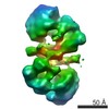 3399C  5fxySC S: Starting model for refinement C: citing same article ( |
|---|---|
| Similar structure data |
-Links
 PDBj
PDBj


- Assembly
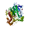
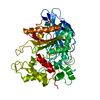
| Deposited unit | 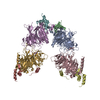
| ||||||||
|---|---|---|---|---|---|---|---|---|---|
| 1 | 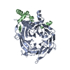
| ||||||||
| 2 | 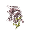
| ||||||||
| 3 | 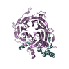
| ||||||||
| 4 | 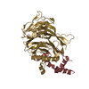
| ||||||||
| Unit cell |
|
-Components
| #1: Protein | Mass: 47709.527 Da / Num. of mol.: 4 Source method: isolated from a genetically manipulated source Source: (gene. exp.) Homo sapiens (human) / Gene: RBBP4, RBAP48 / Plasmid: PCDNA3 / Cell line (production host): HEK293F / Production host: HOMO SAPIENS (human) / References: UniProt: Q09028#2: Protein | Mass: 9773.559 Da / Num. of mol.: 4 Source method: isolated from a genetically manipulated source Source: (gene. exp.) Homo sapiens (human) / Gene: MTA1 / Cell line (production host): HEK293F / Production host: HOMO SAPIENS (human) / References: UniProt: Q13330 |
|---|
-Experimental details
-Experiment
| Experiment | Method: X-RAY DIFFRACTION / Number of used crystals: 5 |
|---|
- Sample preparation
| Crystal | Density Matthews: 2.56 Å3/Da / Density % sol: 52.01 % |
|---|---|
| Crystal grow | Temperature: 298 K / Method: vapor diffusion, sitting drop / pH: 7.5 Details: 0.21 M AMMONIUM CITRATE, 19% PEG 3350, 15 mM MALTOSE pH 7.5 |
-Data collection
| Diffraction | Mean temperature: 100 K |
|---|---|
| Diffraction source | Source: SYNCHROTRON / Site: Diamond / Beamline: I24 / Wavelength: 0.96861 Å |
| Detector | Type: DECTRIS PILATUS3 6M / Detector: PIXEL / Date: Mar 9, 2017 |
| Radiation | Protocol: SINGLE WAVELENGTH / Monochromatic (M) / Laue (L): M / Scattering type: x-ray |
| Radiation wavelength | Wavelength: 0.96861 Å / Relative weight: 1 |
| Reflection | Resolution: 2.8→95.4 Å / Num. obs: 51450 / % possible obs: 95 % / Redundancy: 9.3 % / CC1/2: 0.995 / Rmerge(I) obs: 0.1 / Net I/σ(I): 15.5 |
| Reflection shell | Resolution: 2.8→2.88 Å / Redundancy: 9.2 % / Rmerge(I) obs: 0.82 / Mean I/σ(I) obs: 3.4 / % possible all: 96.9 |
- Processing
| Software |
| ||||||||||||||||||||||||||||||||||||||||||||||||||||||||||||||||||||||||||||||||||||||||||||||||||||||||||||||||||||||||||||||||||||||||||||||||||||||||||||||||||||||||||||||||||||||
|---|---|---|---|---|---|---|---|---|---|---|---|---|---|---|---|---|---|---|---|---|---|---|---|---|---|---|---|---|---|---|---|---|---|---|---|---|---|---|---|---|---|---|---|---|---|---|---|---|---|---|---|---|---|---|---|---|---|---|---|---|---|---|---|---|---|---|---|---|---|---|---|---|---|---|---|---|---|---|---|---|---|---|---|---|---|---|---|---|---|---|---|---|---|---|---|---|---|---|---|---|---|---|---|---|---|---|---|---|---|---|---|---|---|---|---|---|---|---|---|---|---|---|---|---|---|---|---|---|---|---|---|---|---|---|---|---|---|---|---|---|---|---|---|---|---|---|---|---|---|---|---|---|---|---|---|---|---|---|---|---|---|---|---|---|---|---|---|---|---|---|---|---|---|---|---|---|---|---|---|---|---|---|---|
| Refinement | Method to determine structure: MOLECULAR REPLACEMENT Starting model: 5FXY Resolution: 2.8→95.33 Å / Cor.coef. Fo:Fc: 0.922 / Cor.coef. Fo:Fc free: 0.876 / SU B: 17.132 / SU ML: 0.333 / Cross valid method: THROUGHOUT / ESU R Free: 0.407 / Stereochemistry target values: MAXIMUM LIKELIHOOD / Details: HYDROGENS HAVE BEEN ADDED IN THE RIDING POSITIONS
| ||||||||||||||||||||||||||||||||||||||||||||||||||||||||||||||||||||||||||||||||||||||||||||||||||||||||||||||||||||||||||||||||||||||||||||||||||||||||||||||||||||||||||||||||||||||
| Solvent computation | Ion probe radii: 0.8 Å / Shrinkage radii: 0.8 Å / VDW probe radii: 1.2 Å / Solvent model: MASK | ||||||||||||||||||||||||||||||||||||||||||||||||||||||||||||||||||||||||||||||||||||||||||||||||||||||||||||||||||||||||||||||||||||||||||||||||||||||||||||||||||||||||||||||||||||||
| Displacement parameters | Biso mean: 56.404 Å2
| ||||||||||||||||||||||||||||||||||||||||||||||||||||||||||||||||||||||||||||||||||||||||||||||||||||||||||||||||||||||||||||||||||||||||||||||||||||||||||||||||||||||||||||||||||||||
| Refinement step | Cycle: 1 / Resolution: 2.8→95.33 Å
| ||||||||||||||||||||||||||||||||||||||||||||||||||||||||||||||||||||||||||||||||||||||||||||||||||||||||||||||||||||||||||||||||||||||||||||||||||||||||||||||||||||||||||||||||||||||
| Refine LS restraints |
|