 Movie
Movie Controller

[English] 日本語
 Yorodumi
- PDB-3b3w: Crystal structure of the S228A mutant of the aminopeptidase from ... -
+ Open data

- Basic information
| Entry | Database: PDB / ID: 3b3w | ||||||
|---|---|---|---|---|---|---|---|
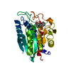
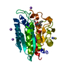
| Title | Crystal structure of the S228A mutant of the aminopeptidase from Vibrio proteolyticus in complex with leucine | ||||||
 Components Components | Bacterial leucyl aminopeptidase | ||||||
 Keywords Keywords | HYDROLASE / alpha beta / Aminopeptidase / Metal-binding / Protease / Secreted / Zinc / Zymogen | ||||||
| Function / homology |  Function and homology information Function and homology informationbacterial leucyl aminopeptidase / metalloexopeptidase activity / aminopeptidase activity / proteolysis / extracellular region / metal ion binding Similarity search - Function | ||||||
| Biological species |  Vibrio proteolyticus (bacteria) Vibrio proteolyticus (bacteria) | ||||||
| Method |  X-RAY DIFFRACTION / SYNCHROTRON / MOLECULAR REPLACEMENT / Resolution: 1.75 Å X-RAY DIFFRACTION / SYNCHROTRON / MOLECULAR REPLACEMENT / Resolution: 1.75 Å | ||||||
 Authors Authors | Ataie, N.J. / Hoang, Q.Q. / Zahniser, M.P.D. / Milne, A. / Petsko, G.A. / Ringe, D. | ||||||
 Citation Citation | Journal: Biochemistry / Year: 2008 Title: Zinc coordination geometry and ligand binding affinity: the structural and kinetic analysis of the second-shell serine 228 residue and the methionine 180 residue of the aminopeptidase from Vibrio proteolyticus. Authors: Ataie, N.J. / Hoang, Q.Q. / Zahniser, M.P. / Tu, Y. / Milne, A. / Petsko, G.A. / Ringe, D. | ||||||
| History |
|
- Structure visualization
| Structure viewer | Molecule: MolmilJmol/JSmol |
|---|
- Downloads & links
-Download
| PDBx/mmCIF format | 3b3w.cif.gz | 76.1 KB | Display | PDBx/mmCIF format |
|---|---|---|---|---|
| PDB format | pdb3b3w.ent.gz | 56.5 KB | Display | PDB format |
| PDBx/mmJSON format | 3b3w.json.gz | Tree view | PDBx/mmJSON format | |
| Others |  Other downloads Other downloads |
-Validation report
| Arichive directory | https://data.pdbj.org/pub/pdb/validation_reports/b3/3b3wftp://data.pdbj.org/pub/pdb/validation_reports/b3/3b3w | HTTPS FTP |
|---|
-Related structure data
| Related structure data |  3b35C 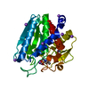 3b3cC  3b3sC  3b3tC  3b3vC 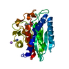 3b7iC  1ampS C: citing same article ( S: Starting model for refinement |
|---|---|
| Similar structure data |


-Links
 PDBj
PDBj


- Assembly
| Deposited unit | 
| ||||||||
|---|---|---|---|---|---|---|---|---|---|
| 1 |
| ||||||||
| Unit cell |
| ||||||||
| Components on special symmetry positions |
| ||||||||
| Details | Authors state that the biological unit of this protein is unknown. |
-Components
-Protein , 1 types, 1 molecules A
| #1: Protein | Mass: 31411.350 Da / Num. of mol.: 1 / Fragment: Residues 107-397 / Mutation: S334A Source method: isolated from a genetically manipulated source Source: (gene. exp.) Vibrio proteolyticus (bacteria) / Strain: DSM 30189 / IFO 13287 / LMG 3772 / NCIMB 1326 / Gene: AAP / Plasmid: pET21b / Species (production host): Escherichia coli / Production host: References: UniProt: Q01693, bacterial leucyl aminopeptidase |
|---|
-Non-polymers , 5 types, 267 molecules 


| #2: Chemical |  Mass: 65.409 Da / Num. of mol.: 2 / Source method: obtained synthetically / Formula: Zn Mass: 65.409 Da / Num. of mol.: 2 / Source method: obtained synthetically / Formula: Zn#3: Chemical | ChemComp-SCN / |  Mass: 58.082 Da / Num. of mol.: 1 / Source method: obtained synthetically / Formula: CNS Mass: 58.082 Da / Num. of mol.: 1 / Source method: obtained synthetically / Formula: CNS#4: Chemical |  Mass: 22.990 Da / Num. of mol.: 2 / Source method: obtained synthetically / Formula: Na Mass: 22.990 Da / Num. of mol.: 2 / Source method: obtained synthetically / Formula: Na#5: Chemical | ChemComp-LEU / |  Type: L-peptide linking / Mass: 131.173 Da / Num. of mol.: 1 / Source method: obtained synthetically / Formula: C6H13NO2 Type: L-peptide linking / Mass: 131.173 Da / Num. of mol.: 1 / Source method: obtained synthetically / Formula: C6H13NO2#6: Water | ChemComp-HOH / | Mass: 18.015 Da / Num. of mol.: 261 / Source method: isolated from a natural source / Formula: H2O |
|---|
-Details
| Has protein modification | Y |
|---|
-Experimental details
-Experiment
| Experiment | Method: X-RAY DIFFRACTION / Number of used crystals: 1 |
|---|
- Sample preparation
| Crystal | Density Matthews: 2.57 Å3/Da / Density % sol: 52.19 % |
|---|---|
| Crystal grow | Temperature: 298 K / Method: vapor diffusion, hanging drop / pH: 8 Details: HEPES, KSCN, NaCl, pH 8.0, VAPOR DIFFUSION, HANGING DROP, temperature 298K |
-Data collection
| Diffraction | Mean temperature: 100 K |
|---|---|
| Diffraction source | Source: SYNCHROTRON / Site: APS  / Beamline: 14-BM-C / Wavelength: 0.9001 Å / Beamline: 14-BM-C / Wavelength: 0.9001 Å |
| Radiation | Protocol: SINGLE WAVELENGTH / Monochromatic (M) / Laue (L): M / Scattering type: x-ray |
| Radiation wavelength | Wavelength: 0.9001 Å / Relative weight: 1 |
| Reflection | Resolution: 1.75→14.98 Å / Num. all: 30654 / Num. obs: 30654 / Observed criterion σ(I): 0 |
- Processing
| Software |
| ||||||||||||||||||||||||||||||||||||||||||||||||||||||||||||||||||||||||||||||||||||||||||||||||||||
|---|---|---|---|---|---|---|---|---|---|---|---|---|---|---|---|---|---|---|---|---|---|---|---|---|---|---|---|---|---|---|---|---|---|---|---|---|---|---|---|---|---|---|---|---|---|---|---|---|---|---|---|---|---|---|---|---|---|---|---|---|---|---|---|---|---|---|---|---|---|---|---|---|---|---|---|---|---|---|---|---|---|---|---|---|---|---|---|---|---|---|---|---|---|---|---|---|---|---|---|---|---|
| Refinement | Method to determine structure: MOLECULAR REPLACEMENT Starting model: PDB entry 1AMP Resolution: 1.75→14.98 Å / Cor.coef. Fo:Fc: 0.962 / Cor.coef. Fo:Fc free: 0.952 / SU B: 2.791 / SU ML: 0.088 / Cross valid method: THROUGHOUT / σ(F): 0 / ESU R: 0.129 / ESU R Free: 0.126 / Stereochemistry target values: MAXIMUM LIKELIHOOD
| ||||||||||||||||||||||||||||||||||||||||||||||||||||||||||||||||||||||||||||||||||||||||||||||||||||
| Solvent computation | Ion probe radii: 0.8 Å / Shrinkage radii: 0.8 Å / VDW probe radii: 1.2 Å / Solvent model: MASK | ||||||||||||||||||||||||||||||||||||||||||||||||||||||||||||||||||||||||||||||||||||||||||||||||||||
| Displacement parameters | Biso mean: 27.352 Å2
| ||||||||||||||||||||||||||||||||||||||||||||||||||||||||||||||||||||||||||||||||||||||||||||||||||||
| Refinement step | Cycle: LAST / Resolution: 1.75→14.98 Å
| ||||||||||||||||||||||||||||||||||||||||||||||||||||||||||||||||||||||||||||||||||||||||||||||||||||
| Refine LS restraints |
| ||||||||||||||||||||||||||||||||||||||||||||||||||||||||||||||||||||||||||||||||||||||||||||||||||||
| LS refinement shell | Resolution: 1.75→1.79 Å / Total num. of bins used: 20
|
