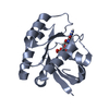
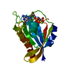
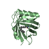
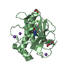
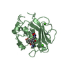
Mass: 18697.680 Da / Num. of mol.: 1 / Fragment: UNP residues 110-219 and 394-450 / Mutation: E108K, Q110V, E121A Source method: isolated from a genetically manipulated source Details: Fusion protein of UNP residues 110-219 and 394-450 from 72 kDa type IV collagenase Source: (gene. exp.) Homo sapiens (human) / Gene: MMP2, CLG4A / Production host: Escherichia coli (E. coli) / Strain (production host): DH5alpha / References: UniProt: P08253, gelatinase A
Method to determine structure: MOLECULAR REPLACEMENT / Resolution: 2→20 Å / Cor.coef. Fo:Fc: 0.954 / Cor.coef. Fo:Fc free: 0.932 / SU B: 6.269 / SU ML: 0.102 / Cross valid method: THROUGHOUT / ESU R Free: 0.158 / Stereochemistry target values: MAXIMUM LIKELIHOOD / Details: HYDROGENS HAVE BEEN USED IF PRESENT IN THE INPUT
Rfactor
Num. reflection
% reflection
Selection details
Rfree
0.20455
578
4.8 %
RANDOM
Rwork
0.16013
-
-
-
obs
0.16229
11476
100 %
-
Solvent computation
Ion probe radii: 0.8 Å / Shrinkage radii: 0.8 Å / VDW probe radii: 1.4 Å / Solvent model: BABINET MODEL WITH MASK
Displacement parameters
Biso mean: 20.375 Å2
Baniso -1
Baniso -2
Baniso -3
1-
-0.05 Å2
0 Å2
0 Å2
2-
-
-0.82 Å2
0 Å2
3-
-
-
0.87 Å2
Refinement step
Cycle: LAST / Resolution: 2→20 Å
Protein
Nucleic acid
Ligand
Solvent
Total
Num. atoms
1392
0
5
263
1660
Refine LS restraints
Refine-ID
Type
Dev ideal
Dev ideal target
Number
X-RAY DIFFRACTION
r_bond_refined_d
0.009
0.021
1438
X-RAY DIFFRACTION
r_bond_other_d
0.001
0.02
963
X-RAY DIFFRACTION
r_angle_refined_deg
1.091
1.937
1954
X-RAY DIFFRACTION
r_angle_other_deg
0.813
3
2324
X-RAY DIFFRACTION
r_dihedral_angle_1_deg
6.121
5
174
X-RAY DIFFRACTION
r_dihedral_angle_2_deg
33.943
23.784
74
X-RAY DIFFRACTION
r_dihedral_angle_3_deg
14.4
15
207
X-RAY DIFFRACTION
r_dihedral_angle_4_deg
11.069
15
7
X-RAY DIFFRACTION
r_chiral_restr
0.07
0.2
193
X-RAY DIFFRACTION
r_gen_planes_refined
0.004
0.021
1650
X-RAY DIFFRACTION
r_gen_planes_other
0.001
0.02
321
X-RAY DIFFRACTION
r_nbd_refined
X-RAY DIFFRACTION
r_nbd_other
X-RAY DIFFRACTION
r_nbtor_refined
X-RAY DIFFRACTION
r_nbtor_other
X-RAY DIFFRACTION
r_xyhbond_nbd_refined
X-RAY DIFFRACTION
r_xyhbond_nbd_other
X-RAY DIFFRACTION
r_metal_ion_refined
X-RAY DIFFRACTION
r_metal_ion_other
X-RAY DIFFRACTION
r_symmetry_vdw_refined
X-RAY DIFFRACTION
r_symmetry_vdw_other
X-RAY DIFFRACTION
r_symmetry_hbond_refined
X-RAY DIFFRACTION
r_symmetry_hbond_other
X-RAY DIFFRACTION
r_symmetry_metal_ion_refined
X-RAY DIFFRACTION
r_symmetry_metal_ion_other
X-RAY DIFFRACTION
r_mcbond_it
X-RAY DIFFRACTION
r_mcbond_other
X-RAY DIFFRACTION
r_mcangle_it
X-RAY DIFFRACTION
r_scbond_it
X-RAY DIFFRACTION
r_scangle_it
X-RAY DIFFRACTION
r_rigid_bond_restr
X-RAY DIFFRACTION
r_sphericity_free
X-RAY DIFFRACTION
r_sphericity_bonded
LS refinement shell
Resolution: 2→2.051 Å / Total num. of bins used: 20
Rfactor
Num. reflection
% reflection
Rfree
0.311
36
-
Rwork
0.171
819
-
obs
-
-
100 %
Refinement TLS params.
Method: refined / Origin x: 1.3733 Å / Origin y: -11.8568 Å / Origin z: -4.699 Å
11
12
13
21
22
23
31
32
33
T
0.0184 Å2
-0.0068 Å2
0.011 Å2
-
0.027 Å2
0.0012 Å2
-
-
0.0106 Å2
L
0.8147 °2
-0.3359 °2
0.0468 °2
-
1.3019 °2
0.0702 °2
-
-
0.6166 °2
S
0.003 Å °
0.0001 Å °
0.028 Å °
-0.032 Å °
0.0356 Å °
-0.0463 Å °
-0.0211 Å °
0.0282 Å °
-0.0386 Å °
+
About Yorodumi
-
News
-
Feb 9, 2022. New format data for meta-information of EMDB entries
New format data for meta-information of EMDB entries
Version 3 of the EMDB header file is now the official format.
The previous official version 1.9 will be removed from the archive.
In the structure databanks used in Yorodumi, some data are registered as the other names, "COVID-19 virus" and "2019-nCoV". Here are the details of the virus and the list of structure data.
Jan 31, 2019. EMDB accession codes are about to change! (news from PDBe EMDB page)
EMDB accession codes are about to change! (news from PDBe EMDB page)
The allocation of 4 digits for EMDB accession codes will soon come to an end. Whilst these codes will remain in use, new EMDB accession codes will include an additional digit and will expand incrementally as the available range of codes is exhausted. The current 4-digit format prefixed with “EMD-” (i.e. EMD-XXXX) will advance to a 5-digit format (i.e. EMD-XXXXX), and so on. It is currently estimated that the 4-digit codes will be depleted around Spring 2019, at which point the 5-digit format will come into force.
The EM Navigator/Yorodumi systems omit the EMD- prefix.
Related info.:Q: What is EMD? / ID/Accession-code notation in Yorodumi/EM Navigator
Yorodumi is a browser for structure data from EMDB, PDB, SASBDB, etc.
This page is also the successor to EM Navigator detail page, and also detail information page/front-end page for Omokage search.
The word "yorodu" (or yorozu) is an old Japanese word meaning "ten thousand". "mi" (miru) is to see.
Related info.:EMDB / PDB / SASBDB / Comparison of 3 databanks / Yorodumi Search / Aug 31, 2016. New EM Navigator & Yorodumi / Yorodumi Papers / Jmol/JSmol / Function and homology information / Changes in new EM Navigator and Yorodumi
 Movie
Movie Controller

 Yorodumi
 Open data
 Basic information
 Components
 Keywords
 Function and homology information
 Homo sapiens (human)
 X-RAY DIFFRACTION /
  Authors
 Citation
 Structure visualization
 Downloads & links
 Other downloads
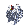


 PDBj
PDBj


 Assembly


 Mass: 65.409 Da / Num. of mol.: 2 / Source method: obtained synthetically / Formula: Zn
 Mass: 40.078 Da / Num. of mol.: 3 / Source method: obtained synthetically / Formula: Ca
 Mass: 18.015 Da / Num. of mol.: 263 / Source method: isolated from a natural source / Formula: H2O
 Sample preparation
 Processing