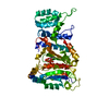
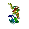
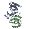
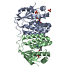
- PDB-3au3: Crystal structure of armadillo repeat domain of APC -
+
Open data
ID or keywords:
Loading...
-
Basic information
Entry
Database: PDB / ID: 3au3
Title
Crystal structure of armadillo repeat domain of APC
Components
Adenomatous polyposis coli protein
Keywords
SIGNALING PROTEIN / Armadillo repeat / signal transduction / Structural Genomics / NPPSFA / National Project on Protein Structural and Functional Analyses / RIKEN Structural Genomics/Proteomics Initiative / RSGI
Function / homology
Function and homology information
APC truncation mutants are not K63 polyubiquitinated / negative regulation of cell cycle G1/S phase transition / negative regulation of cyclin-dependent protein serine/threonine kinase activity / gamma-catenin binding / regulation of attachment of spindle microtubules to kinetochore / positive regulation of pseudopodium assembly / positive regulation of protein localization to centrosome / bicellular tight junction assembly / pattern specification process / negative regulation of microtubule depolymerization ...APC truncation mutants are not K63 polyubiquitinated / negative regulation of cell cycle G1/S phase transition / negative regulation of cyclin-dependent protein serine/threonine kinase activity / gamma-catenin binding / regulation of attachment of spindle microtubules to kinetochore / positive regulation of pseudopodium assembly / positive regulation of protein localization to centrosome / bicellular tight junction assembly / pattern specification process / negative regulation of microtubule depolymerization / heart valve development / beta-catenin destruction complex / catenin complex / APC truncation mutants have impaired AXIN binding / AXIN missense mutants destabilize the destruction complex / Truncations of AMER1 destabilize the destruction complex / microtubule plus-end binding / protein kinase regulator activity / Beta-catenin phosphorylation cascade / Signaling by GSK3beta mutants / CTNNB1 S33 mutants aren't phosphorylated / CTNNB1 S37 mutants aren't phosphorylated / CTNNB1 S45 mutants aren't phosphorylated / CTNNB1 T41 mutants aren't phosphorylated / Wnt signalosome / regulation of microtubule-based process / cell fate specification / Disassembly of the destruction complex and recruitment of AXIN to the membrane / endocardial cushion morphogenesis / Apoptotic cleavage of cellular proteins / negative regulation of G1/S transition of mitotic cell cycle / mitotic spindle assembly checkpoint signaling / regulation of microtubule-based movement / mitotic cytokinesis / dynein complex binding / lateral plasma membrane / bicellular tight junction / adherens junction / Deactivation of the beta-catenin transactivating complex / negative regulation of canonical Wnt signaling pathway / beta-catenin binding / kinetochore / Degradation of beta-catenin by the destruction complex / ruffle membrane / Wnt signaling pathway / positive regulation of protein catabolic process / insulin receptor signaling pathway / Ovarian tumor domain proteases / nervous system development / cell migration / lamellipodium / positive regulation of cold-induced thermogenesis / protein-containing complex assembly / microtubule binding / microtubule / proteasome-mediated ubiquitin-dependent protein catabolic process / cell adhesion / positive regulation of cell migration / positive regulation of apoptotic process / negative regulation of cell population proliferation / DNA damage response / ubiquitin protein ligase binding / centrosome / protein kinase binding / perinuclear region of cytoplasm / Golgi apparatus / nucleoplasm / nucleus / plasma membrane / cytoplasm / cytosol Similarity search - Function
Adenomatous polyposis coli protein repeat / SAMP / EB-1 binding / Adenomatous polyposis coli protein basic domain / Adenomatous polyposis coli protein, 15 residue repeat / Adenomatous polyposis coli (APC) family / Adenomatous polyposis coli protein / Adenomatous polyposis coli, N-terminal dimerisation domain / APC, N-terminal coiled-coil domain superfamily / Adenomatous polyposis coli (APC) repeat ...Adenomatous polyposis coli protein repeat / SAMP / EB-1 binding / Adenomatous polyposis coli protein basic domain / Adenomatous polyposis coli protein, 15 residue repeat / Adenomatous polyposis coli (APC) family / Adenomatous polyposis coli protein / Adenomatous polyposis coli, N-terminal dimerisation domain / APC, N-terminal coiled-coil domain superfamily / Adenomatous polyposis coli (APC) repeat / APC repeat / SAMP Motif / EB-1 Binding Domain / APC basic domain / APC 15 residue motif / Adenomatous polyposis coli tumour suppressor protein / Armadillo-associated region on APC / Unstructured region on APC between 1st and 2nd catenin-bdg motifs / Unstructured region on APC between 1st two creatine-rich regions / Unstructured region on APC between APC_crr and SAMP / Unstructured region on APC between SAMP and APC_crr / Unstructured region on APC between APC_crr regions 5 and 6 / Coiled-coil N-terminus of APC, dimerisation domain / Adenomatous polyposis coli (APC) repeat / Armadillo/plakoglobin ARM repeat profile. / Armadillo/beta-catenin-like repeat / Armadillo/beta-catenin-like repeats / Armadillo / Leucine-rich Repeat Variant / Leucine-rich Repeat Variant / Alpha Horseshoe / Armadillo-like helical / Armadillo-type fold / Mainly Alpha Similarity search - Domain/homology
In the structure databanks used in Yorodumi, some data are registered as the other names, "COVID-19 virus" and "2019-nCoV". Here are the details of the virus and the list of structure data.
Jan 31, 2019. EMDB accession codes are about to change! (news from PDBe EMDB page)
EMDB accession codes are about to change! (news from PDBe EMDB page)
The allocation of 4 digits for EMDB accession codes will soon come to an end. Whilst these codes will remain in use, new EMDB accession codes will include an additional digit and will expand incrementally as the available range of codes is exhausted. The current 4-digit format prefixed with “EMD-” (i.e. EMD-XXXX) will advance to a 5-digit format (i.e. EMD-XXXXX), and so on. It is currently estimated that the 4-digit codes will be depleted around Spring 2019, at which point the 5-digit format will come into force.
The EM Navigator/Yorodumi systems omit the EMD- prefix.
Related info.:Q: What is EMD? / ID/Accession-code notation in Yorodumi/EM Navigator
Yorodumi is a browser for structure data from EMDB, PDB, SASBDB, etc.
This page is also the successor to EM Navigator detail page, and also detail information page/front-end page for Omokage search.
The word "yorodu" (or yorozu) is an old Japanese word meaning "ten thousand". "mi" (miru) is to see.
Related info.:EMDB / PDB / SASBDB / Comparison of 3 databanks / Yorodumi Search / Aug 31, 2016. New EM Navigator & Yorodumi / Yorodumi Papers / Jmol/JSmol / Function and homology information / Changes in new EM Navigator and Yorodumi
 Movie
Movie Controller


 Open data
 Basic information
 Components
 Keywords
 Function and homology information
 Homo sapiens (human)
 X-RAY DIFFRACTION /
  Authors
 Citation
 Structure visualization
 Downloads & links
 Other downloads
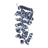


 PDBj
PDBj


 Assembly
 Mass: 18.015 Da / Num. of mol.: 117 / Source method: isolated from a natural source / Formula: H2O
 Sample preparation
 / Beamline: BL26B2 / Wavelength: 0.97897, 0.97934, 0.964
 Processing