 Movie
Movie Controller

[English] 日本語
 Yorodumi
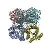
- PDB-3ak4: Crystal structure of NADH-dependent quinuclidinone reductase from... -
+ Open data

- Basic information
| Entry | Database: PDB / ID: 3ak4 | ||||||
|---|---|---|---|---|---|---|---|
| Title | Crystal structure of NADH-dependent quinuclidinone reductase from agrobacterium tumefaciens | ||||||
 Components Components | NADH-dependent quinuclidinone reductase | ||||||
 Keywords Keywords | OXIDOREDUCTASE / SDR / 3-QUINUCLIDINONE / (R)-3-QUINUCLIDINOL / CHIRAL ALCOHOL / CARBONYL REDUCTASE | ||||||
| Function / homology |  Function and homology information Function and homology informationoxidoreductase activity, acting on the CH-OH group of donors, NAD or NADP as acceptor / quinone binding / fatty acid biosynthetic process / nucleotide binding Similarity search - Function | ||||||
| Biological species |  Agrobacterium tumefaciens (bacteria) Agrobacterium tumefaciens (bacteria) | ||||||
| Method |  X-RAY DIFFRACTION / MOLECULAR REPLACEMENT / Resolution: 2 Å X-RAY DIFFRACTION / MOLECULAR REPLACEMENT / Resolution: 2 Å | ||||||
 Authors Authors | Miyakawa, T. / Kataoka, M. / Takeshita, D. / Nomoto, F. / Nagata, K. / Shimizu, S. / Tanokura, M. | ||||||
 Citation Citation | Journal: To be Published Title: Crystal structure of NADH-dependent quinuclidinone reductase from Agrobacterium tumefaciens Authors: Miyakawa, T. / Kataoka, M. / Takeshita, D. / Nomoto, F. / Nagata, K. / Shimizu, S. / Tanokura, M. | ||||||
| History |
|
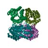
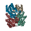
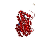
- Structure visualization
| Structure viewer | Molecule: MolmilJmol/JSmol |
|---|
- Downloads & links
-Download
| PDBx/mmCIF format | 3ak4.cif.gz | 216.7 KB | Display | PDBx/mmCIF format |
|---|---|---|---|---|
| PDB format | pdb3ak4.ent.gz | 173.1 KB | Display | PDB format |
| PDBx/mmJSON format | 3ak4.json.gz | Tree view | PDBx/mmJSON format | |
| Others |  Other downloads Other downloads |
-Validation report
| Arichive directory | https://data.pdbj.org/pub/pdb/validation_reports/ak/3ak4ftp://data.pdbj.org/pub/pdb/validation_reports/ak/3ak4 | HTTPS FTP |
|---|
-Related structure data
| Related structure data |  1gegS S: Starting model for refinement |
|---|---|
| Similar structure data |

-Links
 PDBj
PDBj


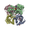
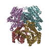
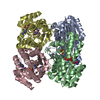
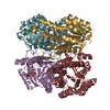
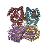
- Assembly
| Deposited unit | 
| ||||||||
|---|---|---|---|---|---|---|---|---|---|
| 1 |
| ||||||||
| Unit cell |
|
-Components
| #1: Protein | Mass: 27588.557 Da / Num. of mol.: 4 Source method: isolated from a genetically manipulated source Source: (gene. exp.) Agrobacterium tumefaciens (bacteria) / Plasmid: PET28A / Production host: #2: Chemical | ChemComp-NAD /   Mass: 663.425 Da / Num. of mol.: 4 / Source method: obtained synthetically / Formula: C21H27N7O14P2 / Comment: NAD*YM Mass: 663.425 Da / Num. of mol.: 4 / Source method: obtained synthetically / Formula: C21H27N7O14P2 / Comment: NAD*YM#3: Water | ChemComp-HOH / |  Mass: 18.015 Da / Num. of mol.: 820 / Source method: isolated from a natural source / Formula: H2O Mass: 18.015 Da / Num. of mol.: 820 / Source method: isolated from a natural source / Formula: H2OSequence details | THE SEQUENCE DATABASE REFERENCE FOR THIS PROTEIN DOES NOT CURRENTLY EXIST. THREE N-TERMINAL ...THE SEQUENCE DATABASE REFERENCE FOR THIS PROTEIN DOES NOT CURRENTLY EXIST. THREE N-TERMINAL RESIDUES ARE EXPRESSION | |
|---|
-Experimental details
-Experiment
| Experiment | Method: X-RAY DIFFRACTION / Number of used crystals: 1 |
|---|
- Sample preparation
| Crystal | Density Matthews: 2.11 Å3/Da / Density % sol: 41.78 % |
|---|---|
| Crystal grow | Temperature: 293 K / Method: vapor diffusion, sitting drop / pH: 8.5 Details: 24% PEG3350, 100MM HEPES, 200MM AMMONIUM ACETATE, pH 8.5, VAPOR DIFFUSION, SITTING DROP, temperature 293K |
-Data collection
| Diffraction | Mean temperature: 95 K |
|---|---|
| Diffraction source | Source: ROTATING ANODE / Type: RIGAKU FR-E DW / Wavelength: 1.5418 / Wavelength: 1.5418 Å |
| Detector | Type: RIGAKU RAXIS VII / Detector: IMAGE PLATE / Date: Aug 3, 2008 |
| Radiation | Monochromator: CONFOCAL MIRROR / Protocol: SINGLE WAVELENGTH / Monochromatic (M) / Laue (L): M / Scattering type: x-ray |
| Radiation wavelength | Wavelength: 1.5418 Å / Relative weight: 1 |
| Reflection | Resolution: 2→20 Å / Num. obs: 61144 / % possible obs: 98.1 % / Redundancy: 3.5 % / Rsym value: 0.045 / Net I/σ(I): 20.6 |
| Reflection shell | Resolution: 2→2.11 Å / Redundancy: 2.9 % / Mean I/σ(I) obs: 7.5 / Num. unique all: 26076 / Rsym value: 0.15 / % possible all: 91.4 |
- Processing
| Software |
| ||||||||||||||||||||||||||||||||||||||||||||||||||||||||||||||||||||||||||||||||||||||||||||||||||||||||||||||||||||||||||||||||||||||||||||||||||||||||||||||||||||||||||
|---|---|---|---|---|---|---|---|---|---|---|---|---|---|---|---|---|---|---|---|---|---|---|---|---|---|---|---|---|---|---|---|---|---|---|---|---|---|---|---|---|---|---|---|---|---|---|---|---|---|---|---|---|---|---|---|---|---|---|---|---|---|---|---|---|---|---|---|---|---|---|---|---|---|---|---|---|---|---|---|---|---|---|---|---|---|---|---|---|---|---|---|---|---|---|---|---|---|---|---|---|---|---|---|---|---|---|---|---|---|---|---|---|---|---|---|---|---|---|---|---|---|---|---|---|---|---|---|---|---|---|---|---|---|---|---|---|---|---|---|---|---|---|---|---|---|---|---|---|---|---|---|---|---|---|---|---|---|---|---|---|---|---|---|---|---|---|---|---|---|---|---|
| Refinement | Method to determine structure: MOLECULAR REPLACEMENT Starting model: PDB ENTRY 1GEG Resolution: 2→20 Å / Cor.coef. Fo:Fc: 0.96 / Cor.coef. Fo:Fc free: 0.938 / SU B: 3.289 / SU ML: 0.094 / Cross valid method: THROUGHOUT / ESU R Free: 0.15 / Stereochemistry target values: MAXIMUM LIKELIHOOD / Details: HYDROGENS HAVE BEEN ADDED IN THE RIDING POSITIONS
| ||||||||||||||||||||||||||||||||||||||||||||||||||||||||||||||||||||||||||||||||||||||||||||||||||||||||||||||||||||||||||||||||||||||||||||||||||||||||||||||||||||||||||
| Solvent computation | Ion probe radii: 0.8 Å / Shrinkage radii: 0.8 Å / VDW probe radii: 1.4 Å / Solvent model: MASK | ||||||||||||||||||||||||||||||||||||||||||||||||||||||||||||||||||||||||||||||||||||||||||||||||||||||||||||||||||||||||||||||||||||||||||||||||||||||||||||||||||||||||||
| Displacement parameters | Biso mean: 15.84 Å2
| ||||||||||||||||||||||||||||||||||||||||||||||||||||||||||||||||||||||||||||||||||||||||||||||||||||||||||||||||||||||||||||||||||||||||||||||||||||||||||||||||||||||||||
| Refinement step | Cycle: LAST / Resolution: 2→20 Å
| ||||||||||||||||||||||||||||||||||||||||||||||||||||||||||||||||||||||||||||||||||||||||||||||||||||||||||||||||||||||||||||||||||||||||||||||||||||||||||||||||||||||||||
| Refine LS restraints |
| ||||||||||||||||||||||||||||||||||||||||||||||||||||||||||||||||||||||||||||||||||||||||||||||||||||||||||||||||||||||||||||||||||||||||||||||||||||||||||||||||||||||||||
| LS refinement shell | Resolution: 2→2.05 Å / Total num. of bins used: 20
|
