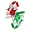
Regulation of ornithine decarboxylase (ODC) / Proteasome assembly / Cross-presentation of soluble exogenous antigens (endosomes) / Autodegradation of Cdh1 by Cdh1:APC/C / APC/C:Cdc20 mediated degradation of Securin / Ubiquitin-Mediated Degradation of Phosphorylated Cdc25A / Ubiquitin-dependent degradation of Cyclin D / AUF1 (hnRNP D0) binds and destabilizes mRNA / Cdc20:Phospho-APC/C mediated degradation of Cyclin A / SCF(Skp2)-mediated degradation of p27/p21 ...Regulation of ornithine decarboxylase (ODC) / Proteasome assembly / Cross-presentation of soluble exogenous antigens (endosomes) / Autodegradation of Cdh1 by Cdh1:APC/C / APC/C:Cdc20 mediated degradation of Securin / Ubiquitin-Mediated Degradation of Phosphorylated Cdc25A / Ubiquitin-dependent degradation of Cyclin D / AUF1 (hnRNP D0) binds and destabilizes mRNA / Cdc20:Phospho-APC/C mediated degradation of Cyclin A / SCF(Skp2)-mediated degradation of p27/p21 / Assembly of the pre-replicative complex / CDK-mediated phosphorylation and removal of Cdc6 / Autodegradation of the E3 ubiquitin ligase COP1 / G2/M Checkpoints / Degradation of AXIN / APC/C:Cdh1 mediated degradation of Cdc20 and other APC/C:Cdh1 targeted proteins in late mitosis/early G1 / Asymmetric localization of PCP proteins / Regulation of RUNX3 expression and activity / Regulation of RAS by GAPs / Regulation of PTEN stability and activity / Regulation of RUNX2 expression and activity / Degradation of GLI1 by the proteasome / UCH proteinases / FBXL7 down-regulates AURKA during mitotic entry and in early mitosis / Degradation of DVL / Orc1 removal from chromatin / GSK3B and BTRC:CUL1-mediated-degradation of NFE2L2 / Dectin-1 mediated noncanonical NF-kB signaling / NIK-->noncanonical NF-kB signaling / TNFR2 non-canonical NF-kB pathway / Hedgehog ligand biogenesis / Hedgehog 'on' state / Oxygen-dependent proline hydroxylation of Hypoxia-inducible Factor Alpha / Degradation of beta-catenin by the destruction complex / Activation of NF-kappaB in B cells / The role of GTSE1 in G2/M progression after G2 checkpoint / FCERI mediated NF-kB activation / CLEC7A (Dectin-1) signaling / Interleukin-1 signaling / RUNX1 regulates transcription of genes involved in differentiation of HSCs / proteasome regulatory particle assembly / Downstream TCR signaling / Separation of Sister Chromatids / MAPK6/MAPK4 signaling / GLI3 is processed to GLI3R by the proteasome / ABC-family proteins mediated transport / Neddylation / positive regulation of cyclin-dependent protein serine/threonine kinase activity / proteasome accessory complex / Ub-specific processing proteases / KEAP1-NFE2L2 pathway / cytosolic proteasome complex / Antigen processing: Ubiquitination & Proteasome degradation / proteasome regulatory particle, base subcomplex / negative regulation of NF-kappaB transcription factor activity / negative regulation of release of cytochrome c from mitochondria / negative regulation of DNA damage response, signal transduction by p53 class mediator / blastocyst development / negative regulation of MAPK cascade / inclusion body / positive regulation of protein ubiquitination / positive regulation of proteasomal ubiquitin-dependent protein catabolic process / microtubule cytoskeleton / positive regulation of cell growth / ciliary basal body / cilium / apoptotic process / negative regulation of apoptotic process / negative regulation of transcription by RNA polymerase II / ATP hydrolysis activity / nucleoplasm / ATP binding / nucleus / cytosol 類似検索 - 分子機能
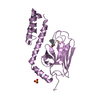
: / : / Proteasomal ATPase OB C-terminal domain / Proteasomal ATPase OB C-terminal domain / Helicase, Ruva Protein; domain 3 - #60 / Ankyrin repeat-containing domain / AAA ATPase, AAA+ lid domain / AAA+ lid domain / ATPase, AAA-type, conserved site / AAA-protein family signature. ...: / : / Proteasomal ATPase OB C-terminal domain / Proteasomal ATPase OB C-terminal domain / Helicase, Ruva Protein; domain 3 - #60 / Ankyrin repeat-containing domain / AAA ATPase, AAA+ lid domain / AAA+ lid domain / ATPase, AAA-type, conserved site / AAA-protein family signature. / Helicase, Ruva Protein; domain 3 / Ankyrin repeat / Ankyrin repeats (3 copies) / Ankyrin repeat profile. / Ankyrin repeat region circular profile. / ATPase family associated with various cellular activities (AAA) / ankyrin repeats / Ankyrin repeat / ATPase, AAA-type, core / Ankyrin repeat-containing domain superfamily / Serine Threonine Protein Phosphatase 5, Tetratricopeptide repeat / Alpha Horseshoe / Nucleic acid-binding, OB-fold / ATPases associated with a variety of cellular activities / AAA+ ATPase domain / P-loop containing nucleoside triphosphate hydrolase / Orthogonal Bundle / Mainly Alpha 類似検索 - ドメイン・相同性
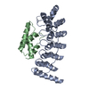
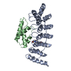
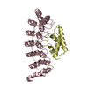
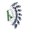
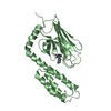
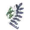
THE 85TH NON-NATURAL RESIDUE IS THE PHOTO REACTED VERSION OF PBPA INCOPORATED BY THE GENETIC CODE ...THE 85TH NON-NATURAL RESIDUE IS THE PHOTO REACTED VERSION OF PBPA INCOPORATED BY THE GENETIC CODE EXPANSION, AND THE 356TH RESIDUE IS THE PHOTO-COVALENT-BONDED PBPA ON GLUTAMIC ACID.
 ムービー
ムービー コントローラー


 データを開く
 基本情報
基本情報 要素
要素 キーワード
キーワード 機能・相同性情報
 X線回折 /
  データ登録者
 引用
引用 構造の表示
構造の表示 ダウンロードとリンク
 その他のダウンロード

 PDBj
PDBj


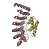
 集合体
集合体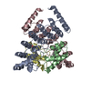

 分子量: 18.015 Da / 分子数: 327 / 由来タイプ: 天然 / 式: H2O
 試料調製
試料調製
 解析
解析