 Movie
Movie Controller

[English] 日本語
 Yorodumi
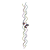
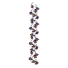
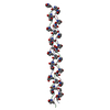
- PDB-3ai6: Triple-helical structure of (D-Pro-D-Pro-Gly)9 at 1.1 A resolution -
+ Open data

- Basic information
| Entry | Database: PDB / ID: 3ai6 | ||||||
|---|---|---|---|---|---|---|---|
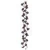
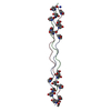
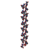
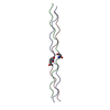
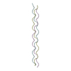
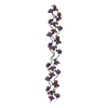
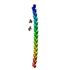
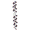
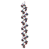
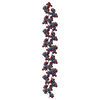
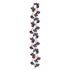
| Title | Triple-helical structure of (D-Pro-D-Pro-Gly)9 at 1.1 A resolution | ||||||
 Components Components | collagen-like peptide | ||||||
 Keywords Keywords | STRUCTURAL PROTEIN / collagen helix / D-enantiomer | ||||||
| Function / homology | Saimiri transformation-associated protein / Collagen triple helix repeat / Collagen triple helix repeat (20 copies) / membrane / Saimiri transformation-associated protein Function and homology information Function and homology information | ||||||
| Method |  X-RAY DIFFRACTION / SYNCHROTRON / MOLECULAR REPLACEMENT / Resolution: 1.1 Å X-RAY DIFFRACTION / SYNCHROTRON / MOLECULAR REPLACEMENT / Resolution: 1.1 Å | ||||||
 Authors Authors | Okuyama, K. / Miyama, K. / Kawaguchi, T. / Nishino, N. | ||||||
 Citation Citation | Journal: To be Published Title: Triple-helical structure of (D-Pro-D-Pro-Gly)9 Authors: Okuyama, K. / Miyama, K. / Kawaguchi, T. / Nishino, N. | ||||||
| History |
|
- Structure visualization
| Structure viewer | Molecule: MolmilJmol/JSmol |
|---|
- Downloads & links
-Download
| PDBx/mmCIF format | 3ai6.cif.gz | 74.7 KB | Display | PDBx/mmCIF format |
|---|---|---|---|---|
| PDB format | pdb3ai6.ent.gz | 66.9 KB | Display | PDB format |
| PDBx/mmJSON format | 3ai6.json.gz | Tree view | PDBx/mmJSON format | |
| Others |  Other downloads Other downloads |
-Validation report
| Summary document | 3ai6_validation.pdf.gz | 444.4 KB | Display | wwPDB validaton report |
|---|---|---|---|---|
| Full document | 3ai6_full_validation.pdf.gz | 444.9 KB | Display | |
| Data in XML | 3ai6_validation.xml.gz | 9.6 KB | Display | |
| Data in CIF | 3ai6_validation.cif.gz | 14.6 KB | Display | |
| Arichive directory | https://data.pdbj.org/pub/pdb/validation_reports/ai/3ai6ftp://data.pdbj.org/pub/pdb/validation_reports/ai/3ai6 | HTTPS FTP |
-Related structure data
| Related structure data | 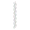 2cuoS S: Starting model for refinement |
|---|---|
| Similar structure data |


-Links
 PDBj
PDBj- Assembly
| Deposited unit | 
| ||||||||
|---|---|---|---|---|---|---|---|---|---|
| 1 | 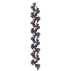
| ||||||||
| 2 | 
| ||||||||
| Unit cell |
|
-Components
| #1: Protein/peptide | Mass: 2279.547 Da / Num. of mol.: 6 / Source method: obtained synthetically Details: This peptide adopts a collagen-helix with opposite chirality. References: UniProt: Q80BK4*PLUS #2: Water | ChemComp-HOH / |  Mass: 18.015 Da / Num. of mol.: 250 / Source method: isolated from a natural source / Formula: H2O Mass: 18.015 Da / Num. of mol.: 250 / Source method: isolated from a natural source / Formula: H2OHas protein modification | N | Sequence details | THE AUTHOR STATES, FOR THE SIX PEPTIDES, THE ACTUAL PEPTIDE CHAIN CONSISTS OF 27 AMINO ACID ...THE AUTHOR STATES, FOR THE SIX PEPTIDES, THE ACTUAL PEPTIDE CHAIN CONSISTS OF 27 AMINO ACID RESIDUES. THE B/F CHAIN CONSISTS OF TWO CHAINS WITH HALF OCCUPANCIE | |
|---|
-Experimental details
-Experiment
| Experiment | Method: X-RAY DIFFRACTION / Number of used crystals: 1 |
|---|
- Sample preparation
| Crystal | Density Matthews: 1.62 Å3/Da / Density % sol: 38.57 % |
|---|---|
| Crystal grow | Temperature: 277 K / Method: vapor diffusion, hanging drop / pH: 5 Details: 11% PEG 200, 0.1M acetate buffer, pH 5.0, VAPOR DIFFUSION, HANGING DROP, temperature 277K |
-Data collection
| Diffraction | Mean temperature: 100 K |
|---|---|
| Diffraction source | Source: SYNCHROTRON / Site: SPring-8  / Beamline: BL44XU / Wavelength: 0.9 Å / Beamline: BL44XU / Wavelength: 0.9 Å |
| Detector | Type: RAYONIX MX225HE / Detector: CCD / Date: Dec 8, 2009 Details: A double-crystal monochromator and a horizontal focusing Mirror |
| Radiation | Monochromator: Mirror / Protocol: SINGLE WAVELENGTH / Monochromatic (M) / Laue (L): M / Scattering type: x-ray |
| Radiation wavelength | Wavelength: 0.9 Å / Relative weight: 1 |
| Reflection twin | Operator: h,-k,-l / Fraction: 0.484 |
| Reflection | Resolution: 1.04→50 Å / Num. obs: 50349 / % possible obs: 96.1 % / Redundancy: 4.7 % / Rmerge(I) obs: 0.051 / Net I/σ(I): 10.19 |
| Reflection shell | Resolution: 1.04→1.06 Å / Redundancy: 3.3 % / Rmerge(I) obs: 0.41 / Mean I/σ(I) obs: 2.72 / Num. unique all: 2337 / % possible all: 89.9 |
- Processing
| Software |
| |||||||||||||||||||||||||||||||||||
|---|---|---|---|---|---|---|---|---|---|---|---|---|---|---|---|---|---|---|---|---|---|---|---|---|---|---|---|---|---|---|---|---|---|---|---|---|
| Refinement | Method to determine structure: MOLECULAR REPLACEMENT Starting model: 2CUO Resolution: 1.1→10 Å / Num. parameters: 11227 / Num. restraintsaints: 14206 / Isotropic thermal model: Anisotropic / Cross valid method: THROUGHOUT / Stereochemistry target values: Engh & Huber Details: The structure was refined under the twinning operator (h, -k, -l) AND TWINNING FRACTION 0.484 using the twinned data.
| |||||||||||||||||||||||||||||||||||
| Refine analyze | Num. disordered residues: 0 / Occupancy sum hydrogen: 0 / Occupancy sum non hydrogen: 1190.5 | |||||||||||||||||||||||||||||||||||
| Refinement step | Cycle: LAST / Resolution: 1.1→10 Å
| |||||||||||||||||||||||||||||||||||
| Refine LS restraints |
| |||||||||||||||||||||||||||||||||||
| LS refinement shell |
|
