 Movie
Movie Controller

[English] 日本語
 Yorodumi
- PDB-3ahw: Crystal Structure of Ustilago sphaerogena Ribonuclease U2 complex... -
+ Open data

- Basic information
| Entry | Database: PDB / ID: 3ahw | ||||||
|---|---|---|---|---|---|---|---|
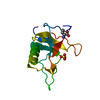
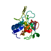
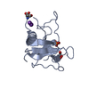
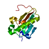
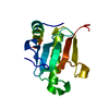
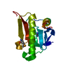
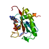
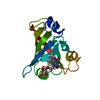
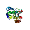
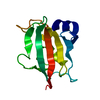
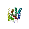
| Title | Crystal Structure of Ustilago sphaerogena Ribonuclease U2 complexed with adenosine 2'-monophosphate | ||||||
 Components Components | Ribonuclease U2 | ||||||
 Keywords Keywords | HYDROLASE / Purine-specific endo-ribonuclease | ||||||
| Function / homology |  Function and homology information Function and homology informationribonuclease U2 / ribonuclease U2 activity / hydrolase activity / RNA binding / metal ion binding Similarity search - Function | ||||||
| Biological species |  Ustilago sphaerogena (fungus) Ustilago sphaerogena (fungus) | ||||||
| Method |  X-RAY DIFFRACTION / SYNCHROTRON / MOLECULAR REPLACEMENT / Resolution: 1.03 Å X-RAY DIFFRACTION / SYNCHROTRON / MOLECULAR REPLACEMENT / Resolution: 1.03 Å | ||||||
 Authors Authors | Noguchi, S. | ||||||
 Citation Citation | Journal: Protein Pept.Lett. / Year: 2010 Title: Conformational variation revealed by the crystal structure of RNase U2A complexed with Ca ion and 2'-adenylic acid at 1.03 angstrom resolution. Authors: Noguchi, S. | ||||||
| History |
|
- Structure visualization
| Structure viewer | Molecule: MolmilJmol/JSmol |
|---|
- Downloads & links
-Download
| PDBx/mmCIF format | 3ahw.cif.gz | 66.8 KB | Display | PDBx/mmCIF format |
|---|---|---|---|---|
| PDB format | pdb3ahw.ent.gz | 48.4 KB | Display | PDB format |
| PDBx/mmJSON format | 3ahw.json.gz | Tree view | PDBx/mmJSON format | |
| Others |  Other downloads Other downloads |
-Validation report
| Arichive directory | https://data.pdbj.org/pub/pdb/validation_reports/ah/3ahwftp://data.pdbj.org/pub/pdb/validation_reports/ah/3ahw | HTTPS FTP |
|---|
-Related structure data
| Related structure data | 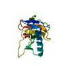 3agnS S: Starting model for refinement |
|---|---|
| Similar structure data |


-Links
 PDBj
PDBj- Assembly
| Deposited unit | 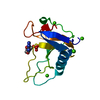
| ||||||||
|---|---|---|---|---|---|---|---|---|---|
| 1 |
| ||||||||
| Unit cell |
|
-Components
| #1: Protein | Mass: 12392.090 Da / Num. of mol.: 1 / Source method: isolated from a natural source / Source: (natural) Ustilago sphaerogena (fungus) / References: UniProt: P00654, EC: 3.1.27.4 | ||||
|---|---|---|---|---|---|
| #2: Chemical | ChemComp-2AM /   Mass: 347.221 Da / Num. of mol.: 1 / Source method: obtained synthetically / Formula: C10H14N5O7P Mass: 347.221 Da / Num. of mol.: 1 / Source method: obtained synthetically / Formula: C10H14N5O7P | ||||
| #3: Chemical | ChemComp-CA /   Mass: 40.078 Da / Num. of mol.: 4 / Source method: obtained synthetically / Formula: Ca Mass: 40.078 Da / Num. of mol.: 4 / Source method: obtained synthetically / Formula: Ca#4: Water | ChemComp-HOH / |  Mass: 18.015 Da / Num. of mol.: 156 / Source method: isolated from a natural source / Formula: H2O Mass: 18.015 Da / Num. of mol.: 156 / Source method: isolated from a natural source / Formula: H2OHas protein modification | Y | |
-Experimental details
-Experiment
| Experiment | Method: X-RAY DIFFRACTION / Number of used crystals: 1 |
|---|
- Sample preparation
| Crystal | Density Matthews: 1.99 Å3/Da / Density % sol: 38.14 % |
|---|---|
| Crystal grow | Temperature: 293 K / Method: hanging drop, vapor diffusion / pH: 6.5 Details: PEG 8000, pH 6.5, hanging drop, vapor diffusion, temperature 293K |
-Data collection
| Diffraction | Mean temperature: 95 K |
|---|---|
| Diffraction source | Source: SYNCHROTRON / Site: Photon Factory  / Beamline: BL-6A / Wavelength: 0.978 Å / Beamline: BL-6A / Wavelength: 0.978 Å |
| Detector | Type: ADSC QUANTUM 4r / Detector: CCD / Date: Jan 25, 2010 |
| Radiation | Monochromator: TRIANGULAR SI(111) / Protocol: SINGLE WAVELENGTH / Monochromatic (M) / Laue (L): M / Scattering type: x-ray |
| Radiation wavelength | Wavelength: 0.978 Å / Relative weight: 1 |
| Reflection | Resolution: 1.03→37.9 Å / Num. all: 48243 / Num. obs: 47871 / % possible obs: 99.2 % / Observed criterion σ(I): 0 / Redundancy: 6.5 % / Biso Wilson estimate: 11.07 Å2 / Rmerge(I) obs: 0.093 / Net I/σ(I): 36.2 |
| Reflection shell | Resolution: 1.03→1.057 Å / Redundancy: 3 % / Rmerge(I) obs: 0.395 / Mean I/σ(I) obs: 2.2 / Num. unique all: 3554 / % possible all: 93.9 |
- Processing
| Software |
| ||||||||||||||||||||||||||||||||||||||||||||||||||||||||||||||||||||||||||||||||||||||||||
|---|---|---|---|---|---|---|---|---|---|---|---|---|---|---|---|---|---|---|---|---|---|---|---|---|---|---|---|---|---|---|---|---|---|---|---|---|---|---|---|---|---|---|---|---|---|---|---|---|---|---|---|---|---|---|---|---|---|---|---|---|---|---|---|---|---|---|---|---|---|---|---|---|---|---|---|---|---|---|---|---|---|---|---|---|---|---|---|---|---|---|---|
| Refinement | Method to determine structure: MOLECULAR REPLACEMENT Starting model: PDB ENTRY 3AGN Resolution: 1.03→25.14 Å / Cor.coef. Fo:Fc: 0.977 / Cor.coef. Fo:Fc free: 0.978 / WRfactor Rfree: 0.137 / WRfactor Rwork: 0.132 / Occupancy max: 1 / Occupancy min: 0.3 / FOM work R set: 0.933 / SU B: 0.592 / SU ML: 0.014 / SU R Cruickshank DPI: 0.025 / SU Rfree: 0.023 / Cross valid method: THROUGHOUT / σ(F): 0 / ESU R: 0.025 / ESU R Free: 0.024 / Stereochemistry target values: MAXIMUM LIKELIHOOD Details: HYDROGENS HAVE BEEN ADDED IN THE RIDING POSITIONS U VALUES: REFINED INDIVIDUALLY
| ||||||||||||||||||||||||||||||||||||||||||||||||||||||||||||||||||||||||||||||||||||||||||
| Solvent computation | Ion probe radii: 0.8 Å / Shrinkage radii: 0.8 Å / VDW probe radii: 1.4 Å / Solvent model: MASK | ||||||||||||||||||||||||||||||||||||||||||||||||||||||||||||||||||||||||||||||||||||||||||
| Displacement parameters | Biso max: 60.02 Å2 / Biso mean: 10.729 Å2 / Biso min: 4.2 Å2
| ||||||||||||||||||||||||||||||||||||||||||||||||||||||||||||||||||||||||||||||||||||||||||
| Refinement step | Cycle: LAST / Resolution: 1.03→25.14 Å
| ||||||||||||||||||||||||||||||||||||||||||||||||||||||||||||||||||||||||||||||||||||||||||
| Refine LS restraints |
| ||||||||||||||||||||||||||||||||||||||||||||||||||||||||||||||||||||||||||||||||||||||||||
| LS refinement shell | Resolution: 1.03→1.057 Å / Total num. of bins used: 20
|
