 Movie
Movie Controller

[English] 日本語
 Yorodumi
- PDB-3agd: Crystal structure of Mglu in its native form in the presence of 4... -
+ Open data

- Basic information
| Entry | Database: PDB / ID: 3agd | ||||||
|---|---|---|---|---|---|---|---|
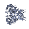
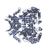
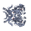
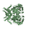
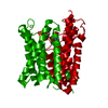
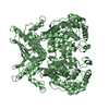
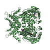
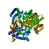
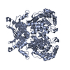
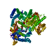
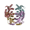
| Title | Crystal structure of Mglu in its native form in the presence of 4.3M NaCl | ||||||
 Components Components | Salt-tolerant glutaminase | ||||||
 Keywords Keywords | HYDROLASE / Glutaminase super family | ||||||
| Function / homology |  Function and homology information Function and homology information | ||||||
| Biological species |  Micrococcus luteus (bacteria) Micrococcus luteus (bacteria) | ||||||
| Method |  X-RAY DIFFRACTION / SYNCHROTRON / MOLECULAR REPLACEMENT / Resolution: 2.2 Å X-RAY DIFFRACTION / SYNCHROTRON / MOLECULAR REPLACEMENT / Resolution: 2.2 Å | ||||||
 Authors Authors | Yoshimune, K. / Shirakihara, Y. / Yumoto, I. | ||||||
 Citation Citation | Journal: To be Published Title: Salt-induced conformational change of salt-tolerant glutaminase from Micrococcus luteus K-3 Authors: Yoshimune, K. / Shirakihara, Y. / Yumoto, I. | ||||||
| History |
|
- Structure visualization
| Structure viewer | Molecule: MolmilJmol/JSmol |
|---|
- Downloads & links
-Download
| PDBx/mmCIF format | 3agd.cif.gz | 187 KB | Display | PDBx/mmCIF format |
|---|---|---|---|---|
| PDB format | pdb3agd.ent.gz | 147.6 KB | Display | PDB format |
| PDBx/mmJSON format | 3agd.json.gz | Tree view | PDBx/mmJSON format | |
| Others |  Other downloads Other downloads |
-Validation report
| Arichive directory | https://data.pdbj.org/pub/pdb/validation_reports/ag/3agdftp://data.pdbj.org/pub/pdb/validation_reports/ag/3agd | HTTPS FTP |
|---|
-Related structure data
| Related structure data | 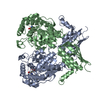 3ageC 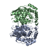 3agfC 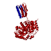 3if5S S: Starting model for refinement C: citing same article ( |
|---|---|
| Similar structure data |


-Links
 PDBj
PDBj
- Assembly
| Deposited unit | 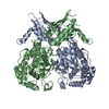
| ||||||||
|---|---|---|---|---|---|---|---|---|---|
| 1 |
| ||||||||
| Unit cell |
|
-Components
| #1: Protein | Mass: 48303.516 Da / Num. of mol.: 2 Source method: isolated from a genetically manipulated source Source: (gene. exp.) Micrococcus luteus (bacteria) / Strain: K-3 / Gene: Glutaminase / Plasmid: pKK223-3 / Production host: #2: Water | ChemComp-HOH / |  Mass: 18.015 Da / Num. of mol.: 770 / Source method: isolated from a natural source / Formula: H2O Mass: 18.015 Da / Num. of mol.: 770 / Source method: isolated from a natural source / Formula: H2O |
|---|
-Experimental details
-Experiment
| Experiment | Method: X-RAY DIFFRACTION / Number of used crystals: 1 |
|---|
- Sample preparation
| Crystal | Density Matthews: 2.95 Å3/Da / Density % sol: 58.31 % |
|---|---|
| Crystal grow | Temperature: 293 K / Method: microbatch method / pH: 7.5 Details: 10mg/ml protein, 50mM HEPES, 1M ammonium formate, 4.3M NaCl, pH 7.5, Microbatch method, temperature 293K |
-Data collection
| Diffraction | Mean temperature: 95 K |
|---|---|
| Diffraction source | Source: SYNCHROTRON / Site: Photon Factory  / Beamline: BL-6A / Wavelength: 0.978 Å / Beamline: BL-6A / Wavelength: 0.978 Å |
| Detector | Type: ADSC QUANTUM 4r / Detector: CCD / Date: Jun 5, 2009 |
| Radiation | Protocol: SINGLE WAVELENGTH / Monochromatic (M) / Laue (L): M / Scattering type: x-ray |
| Radiation wavelength | Wavelength: 0.978 Å / Relative weight: 1 |
| Reflection | Resolution: 2.2→62.3 Å / Num. all: 58860 / Num. obs: 58857 / % possible obs: 100 % / Observed criterion σ(I): 4.8 / Redundancy: 6.8 % / Biso Wilson estimate: 29.2 Å2 / Rmerge(I) obs: 0.09 / Rsym value: 0.09 / Net I/σ(I): 17.3 |
| Reflection shell | Resolution: 2.2→2.32 Å / Redundancy: 6.4 % / Rmerge(I) obs: 0.381 / Mean I/σ(I) obs: 4.8 / Num. unique all: 8473 / Rsym value: 0.381 / % possible all: 100 |
- Processing
| Software |
| |||||||||||||||||||||||||||||||||||||||||||||||||||||||||||||||
|---|---|---|---|---|---|---|---|---|---|---|---|---|---|---|---|---|---|---|---|---|---|---|---|---|---|---|---|---|---|---|---|---|---|---|---|---|---|---|---|---|---|---|---|---|---|---|---|---|---|---|---|---|---|---|---|---|---|---|---|---|---|---|---|---|
| Refinement | Method to determine structure: MOLECULAR REPLACEMENT Starting model: PDB ENTRY 3if5 Resolution: 2.2→59.4 Å / Cross valid method: THROUGHOUT / σ(F): 0
| |||||||||||||||||||||||||||||||||||||||||||||||||||||||||||||||
| Displacement parameters | Biso mean: 29.493 Å2
| |||||||||||||||||||||||||||||||||||||||||||||||||||||||||||||||
| Refine analyze |
| |||||||||||||||||||||||||||||||||||||||||||||||||||||||||||||||
| Refinement step | Cycle: LAST / Resolution: 2.2→59.4 Å
| |||||||||||||||||||||||||||||||||||||||||||||||||||||||||||||||
| Refine LS restraints |
| |||||||||||||||||||||||||||||||||||||||||||||||||||||||||||||||
| LS refinement shell | Refine-ID: X-RAY DIFFRACTION
|
