 Movie
Movie Controller

[English] 日本語
 Yorodumi
- PDB-3ihb: Crystal Structure Analysis of Mglu in its tris and glutamate form -
+ Open data

- Basic information
| Entry | Database: PDB / ID: 3ihb | ||||||
|---|---|---|---|---|---|---|---|
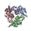
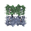
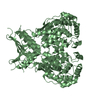
| Title | Crystal Structure Analysis of Mglu in its tris and glutamate form | ||||||
 Components Components | Salt-tolerant glutaminase | ||||||
 Keywords Keywords | HYDROLASE / Salt-tolerant glutaminase | ||||||
| Function / homology |  Function and homology information Function and homology information | ||||||
| Biological species |  Micrococcus luteus (bacteria) Micrococcus luteus (bacteria) | ||||||
| Method |  X-RAY DIFFRACTION / SYNCHROTRON / MOLECULAR REPLACEMENT / molecular replacement / Resolution: 2.4 Å X-RAY DIFFRACTION / SYNCHROTRON / MOLECULAR REPLACEMENT / molecular replacement / Resolution: 2.4 Å | ||||||
 Authors Authors | Yoshimune, K. / Shirakihara, Y. | ||||||
 Citation Citation | Journal: Febs J. / Year: 2010 Title: Crystal structure of salt-tolerant glutaminase from Micrococcus luteus K-3 in the presence and absence of its product l-glutamate and its activator Tris Authors: Yoshimune, K. / Shirakihara, Y. / Wakayama, M. / Yumoto, I. | ||||||
| History |
|
- Structure visualization
| Structure viewer | Molecule: MolmilJmol/JSmol |
|---|
- Downloads & links
-Download
| PDBx/mmCIF format | 3ihb.cif.gz | 188.4 KB | Display | PDBx/mmCIF format |
|---|---|---|---|---|
| PDB format | pdb3ihb.ent.gz | 148.9 KB | Display | PDB format |
| PDBx/mmJSON format | 3ihb.json.gz | Tree view | PDBx/mmJSON format | |
| Others |  Other downloads Other downloads |
-Validation report
| Arichive directory | https://data.pdbj.org/pub/pdb/validation_reports/ih/3ihbftp://data.pdbj.org/pub/pdb/validation_reports/ih/3ihb | HTTPS FTP |
|---|
-Related structure data
| Related structure data |  3if5C  3ih8SC  3ih9C  3ihaC C: citing same article ( S: Starting model for refinement |
|---|---|
| Similar structure data |


-Links
 PDBj
PDBj


- Assembly
| Deposited unit | 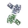
| ||||||||
|---|---|---|---|---|---|---|---|---|---|
| 1 | 
| ||||||||
| 2 | 
| ||||||||
| Unit cell |
|
-Components
| #1: Protein | Mass: 48303.516 Da / Num. of mol.: 2 Source method: isolated from a genetically manipulated source Source: (gene. exp.) Micrococcus luteus (bacteria) / Strain: K-3 / Gene: Glutaminase / Plasmid: pKK223-3 / Production host: #2: Chemical |   Type: L-peptide linking / Mass: 147.129 Da / Num. of mol.: 2 / Source method: obtained synthetically / Formula: C5H9NO4 Type: L-peptide linking / Mass: 147.129 Da / Num. of mol.: 2 / Source method: obtained synthetically / Formula: C5H9NO4#3: Chemical |   Mass: 122.143 Da / Num. of mol.: 2 / Source method: obtained synthetically / Formula: C4H12NO3 / Comment: pH buffer*YM Mass: 122.143 Da / Num. of mol.: 2 / Source method: obtained synthetically / Formula: C4H12NO3 / Comment: pH buffer*YM#4: Water | ChemComp-HOH / |  Mass: 18.015 Da / Num. of mol.: 511 / Source method: isolated from a natural source / Formula: H2O Mass: 18.015 Da / Num. of mol.: 511 / Source method: isolated from a natural source / Formula: H2O |
|---|
-Experimental details
-Experiment
| Experiment | Method: X-RAY DIFFRACTION / Number of used crystals: 1 |
|---|
- Sample preparation
| Crystal | Density Matthews: 3.16 Å3/Da / Density % sol: 61.13 % |
|---|---|
| Crystal grow | Temperature: 293 K / Method: vapor diffusion, hanging drop / pH: 7.2 Details: 15% PEG 4000, 100mM Sodium Acetate, 50mM HEPES, 300mM Tris, 200mM Glutamate, pH 7.2, VAPOR DIFFUSION, HANGING DROP, temperature 293K |
-Data collection
| Diffraction | Mean temperature: 95 K |
|---|---|
| Diffraction source | Source: SYNCHROTRON / Site: Photon Factory  / Beamline: BL-6A / Wavelength: 0.978 Å / Beamline: BL-6A / Wavelength: 0.978 Å |
| Detector | Type: ADSC QUANTUM 4r / Detector: CCD / Date: Feb 20, 2008 |
| Radiation | Protocol: SINGLE WAVELENGTH / Monochromatic (M) / Laue (L): M / Scattering type: x-ray |
| Radiation wavelength | Wavelength: 0.978 Å / Relative weight: 1 |
| Reflection | Resolution: 2.4→57.073 Å / Num. obs: 46819 / % possible obs: 99.5 % / Redundancy: 3.1 % / Rmerge(I) obs: 0.131 / Rsym value: 0.131 / Net I/σ(I): 4.3 |
| Reflection shell | Resolution: 2.4→2.53 Å / Redundancy: 2.8 % / Rmerge(I) obs: 0.412 / Mean I/σ(I) obs: 1.7 / Rsym value: 0.412 / % possible all: 98.6 |
-Phasing
| Phasing | Method: molecular replacement |
|---|
- Processing
| Software |
| ||||||||||||||||||||||||||||
|---|---|---|---|---|---|---|---|---|---|---|---|---|---|---|---|---|---|---|---|---|---|---|---|---|---|---|---|---|---|
| Refinement | Method to determine structure: MOLECULAR REPLACEMENT Starting model: PDB ENTRY 3IH8 Resolution: 2.4→19.86 Å / Rfactor Rfree error: 0.006 / Occupancy max: 1 / Occupancy min: 0.67 / Data cutoff high absF: 2131955 / Data cutoff low absF: 0 / Isotropic thermal model: RESTRAINED / Cross valid method: THROUGHOUT / σ(F): 0 / Details: BULK SOLVENT MODEL USED
| ||||||||||||||||||||||||||||
| Solvent computation | Solvent model: FLAT MODEL / Bsol: 70.711 Å2 / ksol: 0.4 e/Å3 | ||||||||||||||||||||||||||||
| Displacement parameters | Biso max: 116.63 Å2 / Biso mean: 40.938 Å2 / Biso min: 7.6 Å2
| ||||||||||||||||||||||||||||
| Refine analyze |
| ||||||||||||||||||||||||||||
| Refinement step | Cycle: LAST / Resolution: 2.4→19.86 Å
| ||||||||||||||||||||||||||||
| Refine LS restraints |
| ||||||||||||||||||||||||||||
| LS refinement shell | Resolution: 2.4→2.55 Å / Rfactor Rfree error: 0.017 / Total num. of bins used: 6
| ||||||||||||||||||||||||||||
| Xplor file |
|
