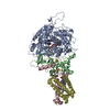
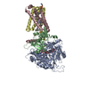
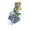
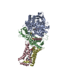
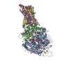
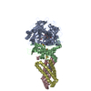
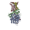
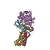
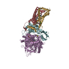
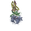
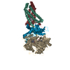
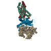
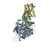
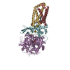
OXIDOREDUCTASE/OXIDOREDUCTASE INHIBITOR / respiratory complex II / inhibitors / Electron transport / Iron / Iron-sulfur / Metal-binding / Mitochondrion / Mitochondrion inner membrane / Oxidoreductase / Transit peptide / Transport / Tricarboxylic acid cycle / Heme / Transmembrane / FAD-binding protein / OXIDOREDUCTASE-OXIDOREDUCTASE INHIBITOR complex
Function / homology
Function and homology information
Maturation of TCA enzymes and regulation of TCA cycle / Citric acid cycle (TCA cycle) / Oxidoreductases; Acting on the CH-OH group of donors; With a quinone or similar compound as acceptor / succinate metabolic process / respiratory chain complex II (succinate dehydrogenase) / mitochondrial electron transport, succinate to ubiquinone / succinate dehydrogenase (quinone) activity / succinate dehydrogenase / 3 iron, 4 sulfur cluster binding / ubiquinone binding ...Maturation of TCA enzymes and regulation of TCA cycle / Citric acid cycle (TCA cycle) / Oxidoreductases; Acting on the CH-OH group of donors; With a quinone or similar compound as acceptor / succinate metabolic process / respiratory chain complex II (succinate dehydrogenase) / mitochondrial electron transport, succinate to ubiquinone / succinate dehydrogenase (quinone) activity / succinate dehydrogenase / 3 iron, 4 sulfur cluster binding / ubiquinone binding / tricarboxylic acid cycle / protein-membrane adaptor activity / aerobic respiration / respiratory electron transport chain / 2 iron, 2 sulfur cluster binding / mitochondrial membrane / flavin adenine dinucleotide binding / nervous system development / 4 iron, 4 sulfur cluster binding / electron transfer activity / mitochondrial inner membrane / mitochondrial matrix / heme binding / metal ion binding Similarity search - Function
Resolution: 2.91→48.24 Å / Cor.coef. Fo:Fc: 0.931 / Cor.coef. Fo:Fc free: 0.906 / WRfactor Rfree: 0.276 / WRfactor Rwork: 0.247 / Occupancy max: 1 / Occupancy min: 1 / FOM work R set: 0.764 / SU B: 34.515 / SU ML: 0.32 / SU R Cruickshank DPI: 0.361 / SU Rfree: 0.417 / Cross valid method: THROUGHOUT / σ(F): 0 / ESU R Free: 0.417 / Stereochemistry target values: MAXIMUM LIKELIHOOD Details: HYDROGENS HAVE BEEN ADDED IN THE RIDING POSITIONS U VALUES : RESIDUAL ONLY
Rfactor
Num. reflection
% reflection
Selection details
Rfree
0.257
1682
5 %
RANDOM
Rwork
0.222
-
-
-
obs
0.223
33975
84.95 %
-
Solvent computation
Ion probe radii: 0.8 Å / Shrinkage radii: 0.8 Å / VDW probe radii: 1.4 Å / Solvent model: MASK
In the structure databanks used in Yorodumi, some data are registered as the other names, "COVID-19 virus" and "2019-nCoV". Here are the details of the virus and the list of structure data.
Jan 31, 2019. EMDB accession codes are about to change! (news from PDBe EMDB page)
EMDB accession codes are about to change! (news from PDBe EMDB page)
The allocation of 4 digits for EMDB accession codes will soon come to an end. Whilst these codes will remain in use, new EMDB accession codes will include an additional digit and will expand incrementally as the available range of codes is exhausted. The current 4-digit format prefixed with “EMD-” (i.e. EMD-XXXX) will advance to a 5-digit format (i.e. EMD-XXXXX), and so on. It is currently estimated that the 4-digit codes will be depleted around Spring 2019, at which point the 5-digit format will come into force.
The EM Navigator/Yorodumi systems omit the EMD- prefix.
Related info.:Q: What is EMD? / ID/Accession-code notation in Yorodumi/EM Navigator
Yorodumi is a browser for structure data from EMDB, PDB, SASBDB, etc.
This page is also the successor to EM Navigator detail page, and also detail information page/front-end page for Omokage search.
The word "yorodu" (or yorozu) is an old Japanese word meaning "ten thousand". "mi" (miru) is to see.
Related info.:EMDB / PDB / SASBDB / Comparison of 3 databanks / Yorodumi Search / Aug 31, 2016. New EM Navigator & Yorodumi / Yorodumi Papers / Jmol/JSmol / Function and homology information / Changes in new EM Navigator and Yorodumi
 Movie
Movie Controller

 Yorodumi
 Open data
 Basic information
 Components
 Keywords
 Function and homology information
 X-RAY DIFFRACTION /
  Authors
 Citation
 Structure visualization
 Downloads & links
 Other downloads
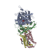


 PDBj
PDBj


 Assembly


 Mass: 785.550 Da / Num. of mol.: 1 / Source method: obtained synthetically / Formula: C27H33N9O15P2 / Comment: FAD*YM
 Mass: 102.046 Da / Num. of mol.: 1 / Source method: obtained synthetically / Formula: C3H2O4
 Mass: 175.820 Da / Num. of mol.: 1 / Source method: obtained synthetically / Formula: Fe2S2
 Mass: 351.640 Da / Num. of mol.: 1 / Source method: obtained synthetically / Formula: Fe4S4
 Mass: 295.795 Da / Num. of mol.: 1 / Source method: obtained synthetically / Formula: Fe3S4
 Mass: 616.487 Da / Num. of mol.: 1 / Source method: obtained synthetically / Formula: C34H32FeN4O4
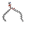
 Mass: 709.933 Da / Num. of mol.: 1 / Source method: obtained synthetically / Formula: C39H68NO8P / Comment: phospholipid*YM
 Mass: 261.060 Da / Num. of mol.: 1 / Source method: obtained synthetically / Formula: C8H8INO
 Sample preparation
 / Beamline: BL-17A / Wavelength: 1 Å
 Processing