 Movie
Movie Controller


+ Open data

- Basic information
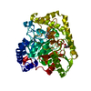
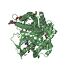
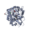
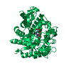
| Entry | Database: PDB / ID: 3a2p | ||||||
|---|---|---|---|---|---|---|---|
| Title | Structure of 6-aminohexanoate cyclic dimer hydrolase | ||||||
 Components Components | 6-aminohexanoate-cyclic-dimer hydrolase | ||||||
 Keywords Keywords | HYDROLASE / alpha/beta fold / Nylon degradation | ||||||
| Function / homology |  Function and homology information Function and homology information6-aminohexanoate-cyclic-dimer hydrolase / 6-aminohexanoate-cyclic-dimer hydrolase activity / nylon catabolic process Similarity search - Function | ||||||
| Biological species |  Arthrobacter sp. (bacteria) Arthrobacter sp. (bacteria) | ||||||
| Method |  X-RAY DIFFRACTION / SYNCHROTRON / SAD / Resolution: 1.9 Å X-RAY DIFFRACTION / SYNCHROTRON / SAD / Resolution: 1.9 Å | ||||||
 Authors Authors | Shibata, N. | ||||||
 Citation Citation | Journal: J.Biol.Chem. / Year: 2010 Title: X-ray crystallographic analysis of the 6-aminohexanoate cyclic dimer hydrolase: catalytic mechanism and evolution of an enzyme responsible for nylon-6 byproduct degradation Authors: Yasuhira, K. / Shibata, N. / Mongami, G. / Uedo, Y. / Atsumi, Y. / Kawashima, Y. / Hibino, A. / Tanaka, Y. / Lee, Y.-H. / Kato, D. / Takeo, M. / Higuchi, Y. / Negoro, S. | ||||||
| History |
|
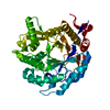
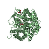
- Structure visualization
| Structure viewer | Molecule: MolmilJmol/JSmol |
|---|
- Downloads & links
-Download
| PDBx/mmCIF format | 3a2p.cif.gz | 111.4 KB | Display | PDBx/mmCIF format |
|---|---|---|---|---|
| PDB format | pdb3a2p.ent.gz | 85.4 KB | Display | PDB format |
| PDBx/mmJSON format | 3a2p.json.gz | Tree view | PDBx/mmJSON format | |
| Others |  Other downloads Other downloads |
-Validation report
| Arichive directory | https://data.pdbj.org/pub/pdb/validation_reports/a2/3a2pftp://data.pdbj.org/pub/pdb/validation_reports/a2/3a2p | HTTPS FTP |
|---|
-Related structure data


-Links
 PDBj
PDBj- Assembly
| Deposited unit | 
| ||||||||
|---|---|---|---|---|---|---|---|---|---|
| 1 |
| ||||||||
| 2 | 
| ||||||||
| Unit cell |
|
-Components
| #1: Protein | Mass: 52262.859 Da / Num. of mol.: 1 Source method: isolated from a genetically manipulated source Source: (gene. exp.) Arthrobacter sp. (bacteria) / Strain: KI72Description: This bacterium had been called Flavobacterium sp. but reassigned to Arthrobacter strain Plasmid: pUC19 / Production host: References: UniProt: P13398, 6-aminohexanoate-cyclic-dimer hydrolase |
|---|---|
| #2: Chemical | ChemComp-GOL /   Mass: 92.094 Da / Num. of mol.: 1 / Source method: obtained synthetically / Formula: C3H8O3 Mass: 92.094 Da / Num. of mol.: 1 / Source method: obtained synthetically / Formula: C3H8O3 |
| #3: Water | ChemComp-HOH /  Mass: 18.015 Da / Num. of mol.: 445 / Source method: isolated from a natural source / Formula: H2O Mass: 18.015 Da / Num. of mol.: 445 / Source method: isolated from a natural source / Formula: H2O |
-Experimental details
-Experiment
| Experiment | Method: X-RAY DIFFRACTION / Number of used crystals: 1 |
|---|
- Sample preparation
| Crystal | Density Matthews: 2.75 Å3/Da / Density % sol: 55.26 % |
|---|---|
| Crystal grow | Temperature: 283 K / Method: vapor diffusion, sitting drop / pH: 8 Details: 1.0M sodium citrate, 25% (v/v) glycerol, 0.1M imidazole buffer pH 8.0, VAPOR DIFFUSION, SITTING DROP, temperature 283K |
-Data collection
| Diffraction | Mean temperature: 100 K |
|---|---|
| Diffraction source | Source: SYNCHROTRON / Site: SPring-8  / Beamline: BL38B1 / Wavelength: 1 Å / Beamline: BL38B1 / Wavelength: 1 Å |
| Detector | Type: RIGAKU JUPITER 210 / Detector: CCD / Date: Dec 18, 2005 |
| Radiation | Protocol: SINGLE WAVELENGTH / Monochromatic (M) / Laue (L): M / Scattering type: x-ray |
| Radiation wavelength | Wavelength: 1 Å / Relative weight: 1 |
| Reflection | Resolution: 1.9→50 Å / Num. all: 44300 / Num. obs: 44300 / % possible obs: 98.6 % / Observed criterion σ(I): -3 / Redundancy: 11 % / Biso Wilson estimate: 26.6 Å2 / Rmerge(I) obs: 0.038 / Net I/σ(I): 69.1 |
| Reflection shell | Resolution: 1.9→1.97 Å / Redundancy: 9 % / Rmerge(I) obs: 0.304 / Mean I/σ(I) obs: 5.1 / Num. unique all: 4021 / % possible all: 90.4 |
- Processing
| Software |
| ||||||||||||||||||||||||||||||||||||||||||||||||||||||||||||||||||||||||||||||||
|---|---|---|---|---|---|---|---|---|---|---|---|---|---|---|---|---|---|---|---|---|---|---|---|---|---|---|---|---|---|---|---|---|---|---|---|---|---|---|---|---|---|---|---|---|---|---|---|---|---|---|---|---|---|---|---|---|---|---|---|---|---|---|---|---|---|---|---|---|---|---|---|---|---|---|---|---|---|---|---|---|---|
| Refinement | Method to determine structure: SAD / Resolution: 1.9→34.49 Å / Rfactor Rfree error: 0.003 / Data cutoff high absF: 2092520.38 / Data cutoff low absF: 0 / Isotropic thermal model: RESTRAINED / Cross valid method: THROUGHOUT / σ(F): 0 / Stereochemistry target values: Engh & Huber / Details: BULK SOLVENT MODEL USED
| ||||||||||||||||||||||||||||||||||||||||||||||||||||||||||||||||||||||||||||||||
| Solvent computation | Solvent model: FLAT MODEL / Bsol: 76.885 Å2 / ksol: 0.4 e/Å3 | ||||||||||||||||||||||||||||||||||||||||||||||||||||||||||||||||||||||||||||||||
| Displacement parameters | Biso mean: 40.2 Å2
| ||||||||||||||||||||||||||||||||||||||||||||||||||||||||||||||||||||||||||||||||
| Refine analyze |
| ||||||||||||||||||||||||||||||||||||||||||||||||||||||||||||||||||||||||||||||||
| Refinement step | Cycle: LAST / Resolution: 1.9→34.49 Å
| ||||||||||||||||||||||||||||||||||||||||||||||||||||||||||||||||||||||||||||||||
| Refine LS restraints |
| ||||||||||||||||||||||||||||||||||||||||||||||||||||||||||||||||||||||||||||||||
| LS refinement shell | Resolution: 1.9→1.97 Å / Rfactor Rfree error: 0.014 / Total num. of bins used: 10
| ||||||||||||||||||||||||||||||||||||||||||||||||||||||||||||||||||||||||||||||||
| Xplor file |
|
