 Movie
Movie Controller


+ Open data

- Basic information
| Entry | Database: PDB / ID: 3a28 | ||||||
|---|---|---|---|---|---|---|---|
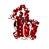
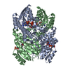
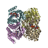
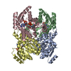
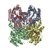
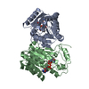
| Title | Crystal structure of L-2,3-butanediol dehydrogenase | ||||||
 Components Components | L-2.3-butanediol dehydrogenase | ||||||
 Keywords Keywords | OXIDOREDUCTASE / chiral substrate recognition | ||||||
| Function / homology |  Function and homology information Function and homology information(S,S)-butanediol dehydrogenase / (S,S)-butanediol dehydrogenase activity / acetoin metabolic process / diacetyl reductase [(S)-acetoin forming] / diacetyl reductase ((S)-acetoin forming) (NAD+) activity / butanediol metabolic process / acetoin catabolic process / NADH binding / NAD+ binding / quinone binding ...(S,S)-butanediol dehydrogenase / (S,S)-butanediol dehydrogenase activity / acetoin metabolic process / diacetyl reductase [(S)-acetoin forming] / diacetyl reductase ((S)-acetoin forming) (NAD+) activity / butanediol metabolic process / acetoin catabolic process / NADH binding / NAD+ binding / quinone binding / fatty acid biosynthetic process / protein homotetramerization Similarity search - Function | ||||||
| Biological species |  Brevibacterium saccharolyticum (bacteria) Brevibacterium saccharolyticum (bacteria) | ||||||
| Method |  X-RAY DIFFRACTION / SYNCHROTRON / MOLECULAR REPLACEMENT / Resolution: 2 Å X-RAY DIFFRACTION / SYNCHROTRON / MOLECULAR REPLACEMENT / Resolution: 2 Å | ||||||
 Authors Authors | Otagiri, M. / Kurisu, G. / Ui, S. / Kusunoki, M. | ||||||
 Citation Citation | Journal: Febs Lett. / Year: 2010 Title: Structural basis for chiral substrate recognition by two 2,3-butanediol dehydrogenases Authors: Otagiri, M. / Ui, S. / Takusagawa, Y. / Ohtsuki, T. / Kurisu, G. / Kusunoki, M. | ||||||
| History |
|
- Structure visualization
| Structure viewer | Molecule: MolmilJmol/JSmol |
|---|
- Downloads & links
-Download
| PDBx/mmCIF format | 3a28.cif.gz | 420.1 KB | Display | PDBx/mmCIF format |
|---|---|---|---|---|
| PDB format | pdb3a28.ent.gz | 343 KB | Display | PDB format |
| PDBx/mmJSON format | 3a28.json.gz | Tree view | PDBx/mmJSON format | |
| Others |  Other downloads Other downloads |
-Validation report
| Summary document | 3a28_validation.pdf.gz | 2.8 MB | Display | wwPDB validaton report |
|---|---|---|---|---|
| Full document | 3a28_full_validation.pdf.gz | 2.9 MB | Display | |
| Data in XML | 3a28_validation.xml.gz | 99.4 KB | Display | |
| Data in CIF | 3a28_validation.cif.gz | 134 KB | Display | |
| Arichive directory | https://data.pdbj.org/pub/pdb/validation_reports/a2/3a28ftp://data.pdbj.org/pub/pdb/validation_reports/a2/3a28 | HTTPS FTP |
-Related structure data
| Related structure data |  1gegS S: Starting model for refinement |
|---|---|
| Similar structure data |


-Links
 PDBj
PDBj


- Assembly
| Deposited unit | 
| ||||||||
|---|---|---|---|---|---|---|---|---|---|
| 1 | 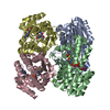
| ||||||||
| 2 | 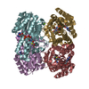
| ||||||||
| Unit cell |
|
-Components
| #1: Protein | Mass: 27128.635 Da / Num. of mol.: 8 Source method: isolated from a genetically manipulated source Source: (gene. exp.) Brevibacterium saccharolyticum (bacteria)Strain: C-1012 / Production host: #2: Chemical | ChemComp-NAD /   Mass: 663.425 Da / Num. of mol.: 8 / Source method: obtained synthetically / Formula: C21H27N7O14P2 / Comment: NAD*YM Mass: 663.425 Da / Num. of mol.: 8 / Source method: obtained synthetically / Formula: C21H27N7O14P2 / Comment: NAD*YM#3: Chemical | ChemComp-BME /   Mass: 78.133 Da / Num. of mol.: 8 / Source method: obtained synthetically / Formula: C2H6OS Mass: 78.133 Da / Num. of mol.: 8 / Source method: obtained synthetically / Formula: C2H6OS#4: Chemical | ChemComp-MG /   Mass: 24.305 Da / Num. of mol.: 4 / Source method: obtained synthetically / Formula: Mg Mass: 24.305 Da / Num. of mol.: 4 / Source method: obtained synthetically / Formula: Mg#5: Water | ChemComp-HOH / |  Mass: 18.015 Da / Num. of mol.: 1554 / Source method: isolated from a natural source / Formula: H2O Mass: 18.015 Da / Num. of mol.: 1554 / Source method: isolated from a natural source / Formula: H2O |
|---|
-Experimental details
-Experiment
| Experiment | Method: X-RAY DIFFRACTION / Number of used crystals: 1 |
|---|
- Sample preparation
| Crystal | Density Matthews: 2.25 Å3/Da / Density % sol: 45.41 % |
|---|---|
| Crystal grow | Method: vapor diffusion, hanging drop / Details: VAPOR DIFFUSION, HANGING DROP |
-Data collection
| Diffraction | Mean temperature: 100 K |
|---|---|
| Diffraction source | Source: SYNCHROTRON / Site: Photon Factory  / Beamline: BL-6A / Wavelength: 1 Å / Beamline: BL-6A / Wavelength: 1 Å |
| Detector | Type: ADSC QUANTUM 4 / Detector: CCD / Date: Dec 18, 2000 |
| Radiation | Protocol: SINGLE WAVELENGTH / Monochromatic (M) / Laue (L): M / Scattering type: x-ray |
| Radiation wavelength | Wavelength: 1 Å / Relative weight: 1 |
| Reflection | Resolution: 2→40 Å / Num. obs: 121123 / % possible obs: 94.4 % / Rmerge(I) obs: 0.062 |
- Processing
| Software |
| ||||||||||||||||||
|---|---|---|---|---|---|---|---|---|---|---|---|---|---|---|---|---|---|---|---|
| Refinement | Method to determine structure: MOLECULAR REPLACEMENT Starting model: PDB ENTRY 1geg Resolution: 2→30 Å / Stereochemistry target values: Engh & Huber /
| ||||||||||||||||||
| Refinement step | Cycle: LAST / Resolution: 2→30 Å
|
