 Movie
Movie Controller

[English] 日本語
 Yorodumi
- PDB-36ja: Coagulation factor V DTQQ (B-domain region 811-1491 truncated, R7... -
+ Open data

- Basic information
| Entry | Database: PDB / ID: 36ja | |||||||||
|---|---|---|---|---|---|---|---|---|---|---|
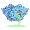
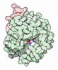
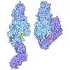
| Title | Coagulation factor V DTQQ (B-domain region 811-1491 truncated, R709Q, R1545Q) in solution-phase | |||||||||
 Components Components | Coagulation factor V | |||||||||
 Keywords Keywords | BLOOD CLOTTING / coagulation factor V / proteinase cofactor / discoidin / solution-phase | |||||||||
| Function / homology |  Function and homology information Function and homology informationresponse to vitamin K / platelet alpha granule / Cargo concentration in the ER / COPII-coated ER to Golgi transport vesicle / COPII-mediated vesicle transport / blood circulation / : / endoplasmic reticulum-Golgi intermediate compartment membrane / platelet alpha granule lumen / Post-translational protein phosphorylation ...response to vitamin K / platelet alpha granule / Cargo concentration in the ER / COPII-coated ER to Golgi transport vesicle / COPII-mediated vesicle transport / blood circulation / : / endoplasmic reticulum-Golgi intermediate compartment membrane / platelet alpha granule lumen / Post-translational protein phosphorylation / blood coagulation / Regulation of Insulin-like Growth Factor (IGF) transport and uptake by Insulin-like Growth Factor Binding Proteins (IGFBPs) / Platelet degranulation / extracellular vesicle / endoplasmic reticulum lumen / copper ion binding / : / extracellular region / membrane / plasma membrane Similarity search - Function | |||||||||
| Biological species |  Homo sapiens (human) Homo sapiens (human) | |||||||||
| Method | ELECTRON MICROSCOPY / single particle reconstruction / cryo EM / Resolution: 3.4 Å | |||||||||
 Authors Authors | Kolyadko, V.N. / Pumroy, R.A. / Moiseenkova-Bell, V.Y. / Krishnaswamy, S. | |||||||||
| Funding support |  United States, 1items United States, 1items
| |||||||||
 Citation Citation | Journal: Proc Natl Acad Sci U S A / Year: 2026 Title: Structural basis for membrane binding by coagulation factors V and VIII and their specificity for phosphatidylserine-containing membranes. Authors: Vladimir N Kolyadko / Ruth A Pumroy / Vera Y Moiseenkova-Bell / Sriram Krishnaswamy / Abstract: Phosphatidylserine on the surface of activated cells serves as a signal for coagulation factor binding and the membrane-dependent assembly of enzyme complexes that drive the accelerated host response ...Phosphatidylserine on the surface of activated cells serves as a signal for coagulation factor binding and the membrane-dependent assembly of enzyme complexes that drive the accelerated host response to vascular damage. Here, we use single-particle cryo-EM of liposome-bound proteins to establish common mechanisms utilized by coagulation factors V and VIII for membrane binding and phospholipid specificity. The interface between these peripheral proteins and the membrane shows contacts between the discoidin C1 and C2 domains and the interfacial phospholipid headgroup region of the membrane bilayer. There is no evidence for the insertion of membrane-contacting protein loops into the membrane core. This refutes previous models of high affinity protein binding by insertion into the hydrophobic core of the membrane. We also resolve density for the headgroup of phosphatidylserine bound to conserved pockets within the C1 and C2 domains. These single binding pockets in each C domain highlight the network of putative hydrogen bonds that contribute to binding specificity for phosphatidylserine. Our results now provide a high-resolution structural view of the protein-membrane interface for these coagulation proteins and highlight the utility of using liposomes as near-physiologic membrane mimetics in structural studies. The principles established in this work may also apply to other biological systems wherein function is regulated by reversible membrane binding. | |||||||||
| History |
|
- Structure visualization
| Structure viewer | Molecule: MolmilJmol/JSmol |
|---|
- Downloads & links
-Download
| PDBx/mmCIF format | 36ja.cif.gz | 259.9 KB | Display | PDBx/mmCIF format |
|---|---|---|---|---|
| PDB format | pdb36ja.ent.gz | 179.2 KB | Display | PDB format |
| PDBx/mmJSON format | 36ja.json.gz | Tree view | PDBx/mmJSON format | |
| Others |  Other downloads Other downloads |
-Validation report
| Arichive directory | https://data.pdbj.org/pub/pdb/validation_reports/6j/36jaftp://data.pdbj.org/pub/pdb/validation_reports/6j/36ja | HTTPS FTP |
|---|
-Related structure data
| Related structure data |  77608MC  36jbC  36jkC  36jlC M: map data used to model this data C: citing same article ( |
|---|---|
| Similar structure data |


-Links
 PDBj
PDBj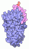



- Assembly
| Deposited unit | 
|
|---|---|
| 1 |
|
-Components
-Protein , 1 types, 1 molecules A
| #1: Protein | Mass: 173755.000 Da / Num. of mol.: 1 / Mutation: R709Q; del811-1491; R1545Q Source method: isolated from a genetically manipulated source Details: Single-chain; B-domain region 811-1491 deleted / Source: (gene. exp.) Homo sapiens (human) / Tissue: blood / Gene: F5 / Cell line (production host): BHK / Production host:  Mesocricetus auratus (golden hamster) / References: UniProt: P12259 Mesocricetus auratus (golden hamster) / References: UniProt: P12259 |
|---|
-Sugars , 3 types, 4 molecules 
| #2: Polysaccharide | 2-acetamido-2-deoxy-beta-D-glucopyranose-(1-4)-2-acetamido-2-deoxy-beta-D-glucopyranose Source method: isolated from a genetically manipulated source |
|---|---|
| #3: Polysaccharide | alpha-D-mannopyranose-(1-3)-[alpha-D-mannopyranose-(1-6)]beta-D-mannopyranose-(1-4)-2-acetamido-2- ...alpha-D-mannopyranose-(1-3)-[alpha-D-mannopyranose-(1-6)]beta-D-mannopyranose-(1-4)-2-acetamido-2-deoxy-beta-D-glucopyranose-(1-4)-2-acetamido-2-deoxy-beta-D-glucopyranose Source method: isolated from a genetically manipulated source |
| #4: Sugar |  Type: D-saccharide, beta linking / Mass: 221.208 Da / Num. of mol.: 2 / Source method: obtained synthetically / Formula: C8H15NO6 Type: D-saccharide, beta linking / Mass: 221.208 Da / Num. of mol.: 2 / Source method: obtained synthetically / Formula: C8H15NO6 |
-Non-polymers , 2 types, 3 molecules 

| #5: Chemical |  Mass: 40.078 Da / Num. of mol.: 2 / Source method: obtained synthetically / Formula: Ca / Feature type: SUBJECT OF INVESTIGATION Mass: 40.078 Da / Num. of mol.: 2 / Source method: obtained synthetically / Formula: Ca / Feature type: SUBJECT OF INVESTIGATION#6: Chemical | ChemComp-CU1 / |  Mass: 63.546 Da / Num. of mol.: 1 / Source method: obtained synthetically / Formula: Cu / Feature type: SUBJECT OF INVESTIGATION Mass: 63.546 Da / Num. of mol.: 1 / Source method: obtained synthetically / Formula: Cu / Feature type: SUBJECT OF INVESTIGATION |
|---|
-Details
| Has ligand of interest | Y |
|---|---|
| Has protein modification | Y |
-Experimental details
-Experiment
| Experiment | Method: ELECTRON MICROSCOPY |
|---|---|
| EM experiment | Aggregation state: PARTICLE / 3D reconstruction method: single particle reconstruction |
- Sample preparation
| Component | Name: Coagulation factor V DTQQ, glycosylated, bound to calcium (II) and copper (I) Type: COMPLEX / Entity ID: #1 / Source: RECOMBINANT | |||||||||||||||||||||||||
|---|---|---|---|---|---|---|---|---|---|---|---|---|---|---|---|---|---|---|---|---|---|---|---|---|---|---|
| Molecular weight | Value: 0.216 MDa / Experimental value: YES | |||||||||||||||||||||||||
| Source (natural) | Organism: Homo sapiens (human) | |||||||||||||||||||||||||
| Source (recombinant) | Organism: Mesocricetus auratus (golden hamster) | |||||||||||||||||||||||||
| Buffer solution | pH: 7.5 | |||||||||||||||||||||||||
| Buffer component |
| |||||||||||||||||||||||||
| Specimen | Conc.: 0.054 mg/ml / Embedding applied: NO / Shadowing applied: NO / Staining applied: NO / Vitrification applied: YES | |||||||||||||||||||||||||
| Specimen support | Grid material: COPPER / Grid mesh size: 300 divisions/in. / Grid type: Quantifoil R0.6/1 | |||||||||||||||||||||||||
| Vitrification | Instrument: FEI VITROBOT MARK IV / Cryogen name: ETHANE / Humidity: 100 % / Chamber temperature: 293 K / Details: double application and blotting |
- Electron microscopy imaging
| Experimental equipment |  Model: Titan Krios / Image courtesy: FEI Company |
|---|---|
| Microscopy | Model: TFS KRIOS |
| Electron gun | Electron source:  FIELD EMISSION GUN / Accelerating voltage: 300 kV / Illumination mode: FLOOD BEAM FIELD EMISSION GUN / Accelerating voltage: 300 kV / Illumination mode: FLOOD BEAM |
| Electron lens | Mode: BRIGHT FIELD / Nominal magnification: 81000 X / Nominal defocus max: 2500 nm / Nominal defocus min: 1000 nm / Cs: 2.7 mm |
| Specimen holder | Cryogen: NITROGEN |
| Image recording | Average exposure time: 3.74 sec. / Electron dose: 48.62 e/Å2 / Film or detector model: GATAN K3 (6k x 4k) / Num. of grids imaged: 1 / Num. of real images: 2703 / Details: 40 frames per movie |
- Processing
| EM software |
| ||||||||||||||||||||||||||||
|---|---|---|---|---|---|---|---|---|---|---|---|---|---|---|---|---|---|---|---|---|---|---|---|---|---|---|---|---|---|
| CTF correction | Type: PHASE FLIPPING AND AMPLITUDE CORRECTION | ||||||||||||||||||||||||||||
| 3D reconstruction | Resolution: 3.4 Å / Resolution method: FSC 0.143 CUT-OFF / Num. of particles: 175078 / Symmetry type: POINT | ||||||||||||||||||||||||||||
| Refinement | Cross valid method: NONE |
