 Movie
Movie Controller

[English] 日本語
 Yorodumi
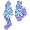
- EMDB-77608: Coagulation factor V DTQQ (B-domain region 811-1491 truncated, R7... -
+ Open data

- Basic information
| Entry |  | |||||||||
|---|---|---|---|---|---|---|---|---|---|---|
| Title | Coagulation factor V DTQQ (B-domain region 811-1491 truncated, R709Q, R1545Q) in solution-phase | |||||||||
 Map data Map data | F5-apo_P20_175K_j024_postprocess.mrc Postprocess B-factor applied -118 A2 | |||||||||
 Sample Sample |
| |||||||||
 Keywords Keywords | coagulation factor V / proteinase cofactor / discoidin / solution-phase / BLOOD CLOTTING | |||||||||
| Function / homology |  Function and homology information Function and homology informationresponse to vitamin K / platelet alpha granule / Cargo concentration in the ER / COPII-coated ER to Golgi transport vesicle / COPII-mediated vesicle transport / blood circulation / : / endoplasmic reticulum-Golgi intermediate compartment membrane / platelet alpha granule lumen / Post-translational protein phosphorylation ...response to vitamin K / platelet alpha granule / Cargo concentration in the ER / COPII-coated ER to Golgi transport vesicle / COPII-mediated vesicle transport / blood circulation / : / endoplasmic reticulum-Golgi intermediate compartment membrane / platelet alpha granule lumen / Post-translational protein phosphorylation / blood coagulation / Regulation of Insulin-like Growth Factor (IGF) transport and uptake by Insulin-like Growth Factor Binding Proteins (IGFBPs) / Platelet degranulation / extracellular vesicle / endoplasmic reticulum lumen / copper ion binding / : / extracellular region / membrane / plasma membrane Similarity search - Function | |||||||||
| Biological species |  Homo sapiens (human) Homo sapiens (human) | |||||||||
| Method | single particle reconstruction / cryo EM / Resolution: 3.4 Å | |||||||||
 Authors Authors | Kolyadko VN / Pumroy RA / Moiseenkova-Bell VY / Krishnaswamy S | |||||||||
| Funding support |  United States, 1 items United States, 1 items
| |||||||||
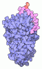
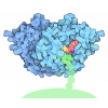
 Citation Citation | Journal: Proc Natl Acad Sci U S A / Year: 2026 Title: Structural basis for membrane binding by coagulation factors V and VIII and their specificity for phosphatidylserine-containing membranes. Authors: Vladimir N Kolyadko / Ruth A Pumroy / Vera Y Moiseenkova-Bell / Sriram Krishnaswamy / Abstract: Phosphatidylserine on the surface of activated cells serves as a signal for coagulation factor binding and the membrane-dependent assembly of enzyme complexes that drive the accelerated host response ...Phosphatidylserine on the surface of activated cells serves as a signal for coagulation factor binding and the membrane-dependent assembly of enzyme complexes that drive the accelerated host response to vascular damage. Here, we use single-particle cryo-EM of liposome-bound proteins to establish common mechanisms utilized by coagulation factors V and VIII for membrane binding and phospholipid specificity. The interface between these peripheral proteins and the membrane shows contacts between the discoidin C1 and C2 domains and the interfacial phospholipid headgroup region of the membrane bilayer. There is no evidence for the insertion of membrane-contacting protein loops into the membrane core. This refutes previous models of high affinity protein binding by insertion into the hydrophobic core of the membrane. We also resolve density for the headgroup of phosphatidylserine bound to conserved pockets within the C1 and C2 domains. These single binding pockets in each C domain highlight the network of putative hydrogen bonds that contribute to binding specificity for phosphatidylserine. Our results now provide a high-resolution structural view of the protein-membrane interface for these coagulation proteins and highlight the utility of using liposomes as near-physiologic membrane mimetics in structural studies. The principles established in this work may also apply to other biological systems wherein function is regulated by reversible membrane binding. | |||||||||
| History |
|
- Structure visualization
| Supplemental images |
|---|

- Downloads & links
-EMDB archive
| Map data | emd_77608.map.gz | 59.9 MB | EMDB map data format | |
|---|---|---|---|---|
| Header (meta data) | emd-77608-v30.xmlemd-77608.xml | 22.8 KB 22.8 KB | Display Display | EMDB header |
| FSC (resolution estimation) | emd_77608_fsc.xml | 9.1 KB | Display | FSC data file |
| Images |  emd_77608.png emd_77608.png | 92.2 KB | ||
| Filedesc metadata | emd-77608.cif.gz | 7.5 KB | ||
| Others | emd_77608_additional_1.map.gzemd_77608_half_map_1.map.gzemd_77608_half_map_2.map.gz | 59.4 MB 49.8 MB 49.7 MB | ||
| Archive directory |  https://data.pdbj.org/pub/emdb/structures/EMD-77608ftp://data.pdbj.org/pub/emdb/structures/EMD-77608 https://data.pdbj.org/pub/emdb/structures/EMD-77608ftp://data.pdbj.org/pub/emdb/structures/EMD-77608 | HTTPS FTP |
-Related structure data
| Related structure data |  36jaMC  36jbC  36jkC  36jlC M: atomic model generated by this map C: citing same article ( |
|---|---|
| Similar structure data |


-Links
| EMDB pages | EMDB (EBI/PDBe) / EMDataResource |
|---|---|
| Related items in Molecule of the Month |



-Map
| File | Download / File: emd_77608.map.gz / Format: CCP4 / Size: 64 MB / Type: IMAGE STORED AS FLOATING POINT NUMBER (4 BYTES) | ||||||||||||||||||||||||||||||||||||
|---|---|---|---|---|---|---|---|---|---|---|---|---|---|---|---|---|---|---|---|---|---|---|---|---|---|---|---|---|---|---|---|---|---|---|---|---|---|
| Annotation | F5-apo_P20_175K_j024_postprocess.mrc Postprocess B-factor applied -118 A2 | ||||||||||||||||||||||||||||||||||||
| Projections & slices | Image control
Images are generated by Spider. | ||||||||||||||||||||||||||||||||||||
| Voxel size | X=Y=Z: 1.07 Å | ||||||||||||||||||||||||||||||||||||
| Density |
| ||||||||||||||||||||||||||||||||||||
| Symmetry | Space group: 1 | ||||||||||||||||||||||||||||||||||||
| Details | EMDB XML:
|
 Z (Sec.)
 Y (Row.)
 X (Col.)


-Supplemental data
-Additional map: F5-apo P20 175K j022 run class001.mrc Unsharpened map
| File | emd_77608_additional_1.map | ||||||||||||
|---|---|---|---|---|---|---|---|---|---|---|---|---|---|
| Annotation | F5-apo_P20_175K_j022_run_class001.mrc Unsharpened map | ||||||||||||
| Projections & Slices |
| ||||||||||||
| Density Histograms |


-Half map: F5apo P20 175K j022 run half1 class001 unfil.mrc half-1
| File | emd_77608_half_map_1.map | ||||||||||||
|---|---|---|---|---|---|---|---|---|---|---|---|---|---|
| Annotation | F5apo_P20_175K_j022_run_half1_class001_unfil.mrc half-1 | ||||||||||||
| Projections & Slices |
| ||||||||||||
| Density Histograms |


-Half map: F5apo P20 175K j022 run half2 class001 unfil.mrc half-2
| File | emd_77608_half_map_2.map | ||||||||||||
|---|---|---|---|---|---|---|---|---|---|---|---|---|---|
| Annotation | F5apo_P20_175K_j022_run_half2_class001_unfil.mrc half-2 | ||||||||||||
| Projections & Slices |
| ||||||||||||
| Density Histograms |


- Sample components
-Entire : Coagulation factor V DTQQ, glycosylated, bound to calcium (II) an...
| Entire | Name: Coagulation factor V DTQQ, glycosylated, bound to calcium (II) and copper (I) |
|---|---|
| Components |
|
-Supramolecule #1: Coagulation factor V DTQQ, glycosylated, bound to calcium (II) an...
| Supramolecule | Name: Coagulation factor V DTQQ, glycosylated, bound to calcium (II) and copper (I) type: complex / ID: 1 / Parent: 0 / Macromolecule list: #1 |
|---|---|
| Source (natural) | Organism: Homo sapiens (human) |
| Molecular weight | Theoretical: 216 KDa |
-Macromolecule #1: Coagulation factor V
| Macromolecule | Name: Coagulation factor V / type: protein_or_peptide / ID: 1 / Details: Single-chain; B-domain region 811-1491 deleted / Number of copies: 1 / Enantiomer: LEVO |
|---|---|
| Source (natural) | Organism: Homo sapiens (human) / Tissue: blood |
| Molecular weight | Theoretical: 173.755 KDa |
| Recombinant expression | Organism:  Mesocricetus auratus (golden hamster) Mesocricetus auratus (golden hamster) |
| Sequence | String: AQLRQFYVAA QGISWSYRPE PTNSSLNLSV TSFKKIVYRE YEPYFKKEKP QSTISGLLGP TLYAEVGDII KVHFKNKADK PLSIHPQGI RYSKLSEGAS YLDHTFPAEK MDDAVAPGRE YTYEWSISED SGPTHDDPPC LTHIYYSHEN LIEDFNSGLI G PLLICKKG ...String: AQLRQFYVAA QGISWSYRPE PTNSSLNLSV TSFKKIVYRE YEPYFKKEKP QSTISGLLGP TLYAEVGDII KVHFKNKADK PLSIHPQGI RYSKLSEGAS YLDHTFPAEK MDDAVAPGRE YTYEWSISED SGPTHDDPPC LTHIYYSHEN LIEDFNSGLI G PLLICKKG TLTEGGTQKT FDKQIVLLFA VFDESKSWSQ SSSLMYTVNG YVNGTMPDIT VCAHDHISWH LLGMSSGPEL FS IHFNGQV LEQNHHKVSA ITLVSATSTT ANMTVGPEGK WIISSLTPKH LQAGMQAYID IKNCPKKTRN LKKITREQRR HMK RWEYFI AAEEVIWDYA PVIPANMDKK YRSQHLDNFS NQIGKHYKKV MYTQYEDESF TKHTVNPNMK EDGILGPIIR AQVR DTLKI VFKNMASRPY SIYPHGVTFS PYEDEVNSSF TSGRNNTMIR AVQPGETYTY KWNILEFDEP TENDAQCLTR PYYSD VDIM RDIASGLIGL LLICKSRSLD RRGIQRAADI EQQAVFAVFD ENKSWYLEDN INKFCENPDE VKRDDPKFYE SNIMST ING YVPESITTLG FCFDDTVQWH FCSVGTQNEI LTIHFTGHSF IYGKRHEDTL TLFPMRGESV TVTMDNVGTW MLTSMNS SP RSKKLRLKFR DVKCIPDDDE DSYEIFEPPE STVMATRKMH DRLEPEDEES DADYDYQNRL AAALGIQSFR NSSLNQEE E EFNLTALALE NGTEFVSSNT DIIVGSNYSS PSNISKFTVN NLAEPQKAPS HQQATTAGSP LRHLIGKNSV LNSSTAEHS SPYSEDPIED TDYIEIIPKE EVQSSEDDYA EIDYVPYDDP YKTDVRTNIN SSRDPDNIAA WYLQSNNGNR RNYYIAAEEI SWDYSEFVQ RETDIEDSDD IPEDTTYKKV VFRKYLDSTF TKRDPRGEYE EHLGILGPII RAEVDDVIQV RFKNLASRPY S LHAHGLSY EKSSEGKTYE DDSPEWFKED NAVQPNSSYT YVWHATERSG PESPGSACRA WAYYSAVNPE KDIHSGLIGP LL ICQKGIL HKDSNMPVDM REFVLLFMTF DEKKSWYYEK KSRSSWRLTS SEMKKSHEFH AINGMIYSLP GLKMYEQEWV RLH LLNIGG SQDIHVVHFH GQTLLENGNK QHQLGVWPLL PGSFKTLEMK ASKPGWWLLN TEVGENQRAG MQTPFLIMDR DCRM PMGLS TGIISDSQIK ASEFLGYWEP RLARLNNGGS YNAWSVEKLA AEFASKPWIQ VDMQKEVIIT GIQTQGAKHY LKSCY TTEF YVAYSSNQIN WQIFKGNSTR NVMYFNGNSD ASTIKENQFD PPIVARYIRI SPTRAYNRPT LRLELQGCEV NGCSTP LGM ENGKIENKQI TASSFKKSWW GDYWEPFRAR LNAQGRVNAW QAKANNNKQW LEIDLLKIKK ITAIITQGCK SLSSEMY VK SYTIHYSEQG VEWKPYRLKS SMVDKIFEGN TNTKGHVKNF FNPPIISRFI RVIPKTWNQS ITLRLELFGC DIY UniProtKB: Coagulation factor V |
-Macromolecule #4: 2-acetamido-2-deoxy-beta-D-glucopyranose
| Macromolecule | Name: 2-acetamido-2-deoxy-beta-D-glucopyranose / type: ligand / ID: 4 / Number of copies: 2 / Formula: NAG |
|---|---|
| Molecular weight | Theoretical: 221.208 Da |
| Chemical component information |  ChemComp-NAG: |
-Macromolecule #5: CALCIUM ION
| Macromolecule | Name: CALCIUM ION / type: ligand / ID: 5 / Number of copies: 2 / Formula: CA |
|---|---|
| Molecular weight | Theoretical: 40.078 Da |
-Macromolecule #6: COPPER (I) ION
| Macromolecule | Name: COPPER (I) ION / type: ligand / ID: 6 / Number of copies: 1 / Formula: CU1 |
|---|---|
| Molecular weight | Theoretical: 63.546 Da |
| Chemical component information |  ChemComp-CU1: |
-Experimental details
-Structure determination
| Method | cryo EM |
|---|---|
 Processing Processing | single particle reconstruction |
| Aggregation state | particle |
-Sample preparation
| Concentration | 0.054 mg/mL | |||||||||||||||
|---|---|---|---|---|---|---|---|---|---|---|---|---|---|---|---|---|
| Buffer | pH: 7.5 Component:
| |||||||||||||||
| Grid | Model: Quantifoil R0.6/1 / Material: COPPER / Mesh: 300 / Support film - Material: CARBON / Support film - topology: HOLEY / Pretreatment - Type: GLOW DISCHARGE / Pretreatment - Time: 40 sec. / Pretreatment - Atmosphere: AIR | |||||||||||||||
| Vitrification | Cryogen name: ETHANE / Chamber humidity: 100 % / Chamber temperature: 293 K / Instrument: FEI VITROBOT MARK IV / Details: double application and blotting. |
- Electron microscopy
| Microscope | TFS KRIOS |
|---|---|
| Image recording | Film or detector model: GATAN K3 (6k x 4k) / Number grids imaged: 1 / Number real images: 2703 / Average exposure time: 3.74 sec. / Average electron dose: 48.62 e/Å2 / Details: 40 frames per movie |
| Electron beam | Acceleration voltage: 300 kV / Electron source:  FIELD EMISSION GUN FIELD EMISSION GUN |
| Electron optics | Illumination mode: FLOOD BEAM / Imaging mode: BRIGHT FIELD / Cs: 2.7 mm / Nominal defocus max: 2.5 µm / Nominal defocus min: 1.0 µm / Nominal magnification: 81000 |
| Sample stage | Cooling holder cryogen: NITROGEN |
| Experimental equipment |  Model: Titan Krios / Image courtesy: FEI Company |

