 Movie
Movie Controller

[English] 日本語
 Yorodumi
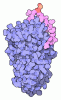
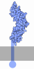
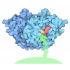
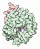
- EMDB-77611: Coagulation factor VIII, full-length, membrane-bound, on Ptd-chol... -
+ Open data

- Basic information
| Entry |  | |||||||||
|---|---|---|---|---|---|---|---|---|---|---|
| Title | Coagulation factor VIII, full-length, membrane-bound, on Ptd-choline : Ptd-serine 75:25 vesicles | |||||||||
 Map data Map data | F8-vesicle_VK02-1_j049_postprocess.mrc Post-processed map. B-factor applied -56 A2 | |||||||||
 Sample Sample |
| |||||||||
 Keywords Keywords | coagulation factor VIII / discoidin / phosphatidylserine / liposome / MEMBRANE PROTEIN | |||||||||
| Function / homology |  Function and homology information Function and homology informationDefective F8 accelerates dissociation of the A2 domain / Defective F8 binding to the cell membrane / Defective F8 secretion / Defective F8 sulfation at Y1699 / Gamma carboxylation, hypusinylation, hydroxylation, and arylsulfatase activation / Defective F8 binding to von Willebrand factor / blood coagulation, intrinsic pathway / Initiation of coagulation cascade / Amplification and propagation of coagulation cascade / Regulation of clotting cascade ...Defective F8 accelerates dissociation of the A2 domain / Defective F8 binding to the cell membrane / Defective F8 secretion / Defective F8 sulfation at Y1699 / Gamma carboxylation, hypusinylation, hydroxylation, and arylsulfatase activation / Defective F8 binding to von Willebrand factor / blood coagulation, intrinsic pathway / Initiation of coagulation cascade / Amplification and propagation of coagulation cascade / Regulation of clotting cascade / Cargo concentration in the ER / COPII-coated ER to Golgi transport vesicle / Defective factor IX causes thrombophilia / Defective cofactor function of FVIIIa variant / Defective F9 variant does not activate FX / COPII-mediated vesicle transport / Defective F8 cleavage by thrombin / endoplasmic reticulum-Golgi intermediate compartment membrane / platelet alpha granule lumen / Golgi lumen / blood coagulation / Platelet degranulation / oxidoreductase activity / endoplasmic reticulum lumen / copper ion binding / extracellular region / plasma membrane Similarity search - Function | |||||||||
| Biological species |  Homo sapiens (human) Homo sapiens (human) | |||||||||
| Method | single particle reconstruction / cryo EM / Resolution: 3.15 Å | |||||||||
 Authors Authors | Kolyadko VN / Pumroy RA / Moiseenkova-Bell VY / Krishnaswamy S | |||||||||
| Funding support |  United States, 1 items United States, 1 items
| |||||||||
 Citation Citation | Journal: Proc Natl Acad Sci U S A / Year: 2026 Title: Structural basis for membrane binding by coagulation factors V and VIII and their specificity for phosphatidylserine-containing membranes. Authors: Vladimir N Kolyadko / Ruth A Pumroy / Vera Y Moiseenkova-Bell / Sriram Krishnaswamy / Abstract: Phosphatidylserine on the surface of activated cells serves as a signal for coagulation factor binding and the membrane-dependent assembly of enzyme complexes that drive the accelerated host response ...Phosphatidylserine on the surface of activated cells serves as a signal for coagulation factor binding and the membrane-dependent assembly of enzyme complexes that drive the accelerated host response to vascular damage. Here, we use single-particle cryo-EM of liposome-bound proteins to establish common mechanisms utilized by coagulation factors V and VIII for membrane binding and phospholipid specificity. The interface between these peripheral proteins and the membrane shows contacts between the discoidin C1 and C2 domains and the interfacial phospholipid headgroup region of the membrane bilayer. There is no evidence for the insertion of membrane-contacting protein loops into the membrane core. This refutes previous models of high affinity protein binding by insertion into the hydrophobic core of the membrane. We also resolve density for the headgroup of phosphatidylserine bound to conserved pockets within the C1 and C2 domains. These single binding pockets in each C domain highlight the network of putative hydrogen bonds that contribute to binding specificity for phosphatidylserine. Our results now provide a high-resolution structural view of the protein-membrane interface for these coagulation proteins and highlight the utility of using liposomes as near-physiologic membrane mimetics in structural studies. The principles established in this work may also apply to other biological systems wherein function is regulated by reversible membrane binding. | |||||||||
| History |
|
- Structure visualization
| Supplemental images |
|---|

- Downloads & links
-EMDB archive
| Map data | emd_77611.map.gz | 85.3 MB | EMDB map data format | |
|---|---|---|---|---|
| Header (meta data) | emd-77611-v30.xmlemd-77611.xml | 25 KB 25 KB | Display Display | EMDB header |
| FSC (resolution estimation) | emd_77611_fsc.xml | 10.3 KB | Display | FSC data file |
| Images |  emd_77611.png emd_77611.png | 123.6 KB | ||
| Filedesc metadata | emd-77611.cif.gz | 8.3 KB | ||
| Others | emd_77611_additional_1.map.gzemd_77611_half_map_1.map.gzemd_77611_half_map_2.map.gz | 83.7 MB 71.2 MB 71.2 MB | ||
| Archive directory |  https://data.pdbj.org/pub/emdb/structures/EMD-77611ftp://data.pdbj.org/pub/emdb/structures/EMD-77611 https://data.pdbj.org/pub/emdb/structures/EMD-77611ftp://data.pdbj.org/pub/emdb/structures/EMD-77611 | HTTPS FTP |
-Related structure data
| Related structure data |  36jlMC  36jaC  36jbC  36jkC M: atomic model generated by this map C: citing same article ( |
|---|---|
| Similar structure data |


-Links
| EMDB pages | EMDB (EBI/PDBe) / EMDataResource |
|---|---|
| Related items in Molecule of the Month |


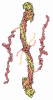
-Map
| File | Download / File: emd_77611.map.gz / Format: CCP4 / Size: 91.1 MB / Type: IMAGE STORED AS FLOATING POINT NUMBER (4 BYTES) | ||||||||||||||||||||||||||||||||||||
|---|---|---|---|---|---|---|---|---|---|---|---|---|---|---|---|---|---|---|---|---|---|---|---|---|---|---|---|---|---|---|---|---|---|---|---|---|---|
| Annotation | F8-vesicle_VK02-1_j049_postprocess.mrc Post-processed map. B-factor applied -56 A2 | ||||||||||||||||||||||||||||||||||||
| Projections & slices | Image control
Images are generated by Spider. | ||||||||||||||||||||||||||||||||||||
| Voxel size | X=Y=Z: 0.887 Å | ||||||||||||||||||||||||||||||||||||
| Density |
| ||||||||||||||||||||||||||||||||||||
| Symmetry | Space group: 1 | ||||||||||||||||||||||||||||||||||||
| Details | EMDB XML:
|
 Z (Sec.)
 Y (Row.)
 X (Col.)


-Supplemental data
-Additional map: j048 run class001.mrc Unsharpened map
| File | emd_77611_additional_1.map | ||||||||||||
|---|---|---|---|---|---|---|---|---|---|---|---|---|---|
| Annotation | j048_run_class001.mrc Unsharpened map | ||||||||||||
| Projections & Slices |
| ||||||||||||
| Density Histograms |


-Half map: j048 run half2 class001 unfil.mrc half-2
| File | emd_77611_half_map_1.map | ||||||||||||
|---|---|---|---|---|---|---|---|---|---|---|---|---|---|
| Annotation | j048_run_half2_class001_unfil.mrc half-2 | ||||||||||||
| Projections & Slices |
| ||||||||||||
| Density Histograms |


-Half map: j048 run half1 class001 unfil.mrc half-1
| File | emd_77611_half_map_2.map | ||||||||||||
|---|---|---|---|---|---|---|---|---|---|---|---|---|---|
| Annotation | j048_run_half1_class001_unfil.mrc half-1 | ||||||||||||
| Projections & Slices |
| ||||||||||||
| Density Histograms |


- Sample components
-Entire : Coagulation factor VIII, glycosylated, bound to calcium (II) and ...
| Entire | Name: Coagulation factor VIII, glycosylated, bound to calcium (II) and copper (I), and to phosphatidyl-L-serine headgroups in small unilamellar vesicles |
|---|---|
| Components |
|
-Supramolecule #1: Coagulation factor VIII, glycosylated, bound to calcium (II) and ...
| Supramolecule | Name: Coagulation factor VIII, glycosylated, bound to calcium (II) and copper (I), and to phosphatidyl-L-serine headgroups in small unilamellar vesicles type: complex / ID: 1 / Parent: 0 / Macromolecule list: #1 Details: Small unilamellar vesicles are composed of a mixture of phospholipids (phosphatidylcholine : phosphatidylserine 75:25) |
|---|---|
| Source (natural) | Organism: Homo sapiens (human) |
| Molecular weight | Theoretical: 330 KDa |
-Macromolecule #1: Coagulation factor VIII
| Macromolecule | Name: Coagulation factor VIII / type: protein_or_peptide / ID: 1 / Number of copies: 1 / Enantiomer: LEVO |
|---|---|
| Source (natural) | Organism: Homo sapiens (human) |
| Molecular weight | Theoretical: 265.061062 KDa |
| Recombinant expression | Organism:  Mesocricetus auratus (golden hamster) Mesocricetus auratus (golden hamster) |
| Sequence | String: ATRRYYLGAV ELSWDYMQSD LGELPVDARF PPRVPKSFPF NTSVVYKKTL FVEFTDHLFN IAKPRPPWMG LLGPTIQAEV YDTVVITLK NMASHPVSLH AVGVSYWKAS EGAEYDDQTS QREKEDDKVF PGGSHTYVWQ VLKENGPMAS DPLCLTYSYL S HVDLVKDL ...String: ATRRYYLGAV ELSWDYMQSD LGELPVDARF PPRVPKSFPF NTSVVYKKTL FVEFTDHLFN IAKPRPPWMG LLGPTIQAEV YDTVVITLK NMASHPVSLH AVGVSYWKAS EGAEYDDQTS QREKEDDKVF PGGSHTYVWQ VLKENGPMAS DPLCLTYSYL S HVDLVKDL NSGLIGALLV CREGSLAKEK TQTLHKFILL FAVFDEGKSW HSETKNSLMQ DRDAASARAW PKMHTVNGYV NR SLPGLIG CHRKSVYWHV IGMGTTPEVH SIFLEGHTFL VRNHRQASLE ISPITFLTAQ TLLMDLGQFL LFCHISSHQH DGM EAYVKV DSCPEEPQLR MKNNEEAEDY DDDLTDSEMD VVRFDDDNSP SFIQIRSVAK KHPKTWVHYI AAEEEDWDYA PLVL APDDR SYKSQYLNNG PQRIGRKYKK VRFMAYTDET FKTREAIQHE SGILGPLLYG EVGDTLLIIF KNQASRPYNI YPHGI TDVR PLYSRRLPKG VKHLKDFPIL PGEIFKYKWT VTVEDGPTKS DPRCLTRYYS SFVNMERDLA SGLIGPLLIC YKESVD QRG NQIMSDKRNV ILFSVFDENR SWYLTENIQR FLPNPAGVQL EDPEFQASNI MHSINGYVFD SLQLSVCLHE VAYWYIL SI GAQTDFLSVF FSGYTFKHKM VYEDTLTLFP FSGETVFMSM ENPGLWILGC HNSDFRNRGM TALLKVSSCD KNTGDYYE D SYEDISAYLL SKNNAIEPRS FSQNSRHPST RQKQFNATTI PENDIEKTDP WFAHRTPMPK IQNVSSSDLL MLLRQSPTP HGLSLSDLQE AKYETFSDDP SPGAIDSNNS LSEMTHFRPQ LHHSGDMVFT PESGLQLRLN EKLGTTAATE LKKLDFKVSS TSNNLISTI PSDNLAAGTD NTSSLGPPSM PVHYDSQLDT TLFGKKSSPL TESGGPLSLS EENNDSKLLE SGLMNSQESS W GKNVSSTE SGRLFKGKRA HGPALLTKDN ALFKVSISLL KTNKTSNNSA TNRKTHIDGP SLLIENSPSV WQNILESDTE FK KVTPLIH DRMLMDKNAT ALRLNHMSNK TTSSKNMEMV QQKKEGPIPP DAQNPDMSFF KMLFLPESAR WIQRTHGKNS LNS GQGPSP KQLVSLGPEK SVEGQNFLSE KNKVVVGKGE FTKDVGLKEM VFPSSRNLFL TNLDNLHENN THNQEKKIQE EIEK KETLI QENVVLPQIH TVTGTKNFMK NLFLLSTRQN VEGSYDGAYA PVLQDFRSLN DSTNRTKKHT AHFSKKGEEE NLEGL GNQT KQIVEKYACT TRISPNTSQQ NFVTQRSKRA LKQFRLPLEE TELEKRIIVD DTSTQWSKNM KHLTPSTLTQ IDYNEK EKG AITQSPLSDC LTRSHSIPQA NRSPLPIAKV SSFPSIRPIY LTRVLFQDNS SHLPAASYRK KDSGVQESSH FLQGAKK NN LSLAILTLEM TGDQREVGSL GTSATNSVTY KKVENTVLPK PDLPKTSGKV ELLPKVHIYQ KDLFPTETSN GSPGHLDL V EGSLLQGTEG AIKWNEANRP GKVPFLRVAT ESSAKTPSKL LDPLAWDNHY GTQIPKEEWK SQEKSPEKTA FKKKDTILS LNACESNHAI AAINEGQNKP EIEVTWAKQG RTERLCSQNP PVLKRHQREI TRTTLQSDQE EIDYDDTISV EMKKEDFDIY DEDENQSPR SFQKKTRHYF IAAVERLWDY GMSSSPHVLR NRAQSGSVPQ FKKVVFQEFT DGSFTQPLYR GELNEHLGLL G PYIRAEVE DNIMVTFRNQ ASRPYSFYSS LISYEEDQRQ GAEPRKNFVK PNETKTYFWK VQHHMAPTKD EFDCKAWAYF SD VDLEKDV HSGLIGPLLV CHTNTLNPAH GRQVTVQEFA LFFTIFDETK SWYFTENMER NCRAPCNIQM EDPTFKENYR FHA INGYIM DTLPGLVMAQ DQRIRWYLLS MGSNENIHSI HFSGHVFTVR KKEEYKMALY NLYPGVFETV EMLPSKAGIW RVEC LIGEH LHAGMSTLFL VYSNKCQTPL GMASGHIRDF QITASGQYGQ WAPKLARLHY SGSINAWSTK EPFSWIKVDL LAPMI IHGI KTQGARQKFS SLYISQFIIM YSLDGKKWQT YRGNSTGTLM VFFGNVDSSG IKHNIFNPPI IARYIRLHPT HYSIRS TLR MELMGCDLNS CSMPLGMESK AISDAQITAS SYFTNMFATW SPSKARLHLQ GRSNAWRPQV NNPKEWLQVD FQKTMKV TG VTTQGVKSLL TSMYVKEFLI SSSQDGHQWT LFFQNGKVKV FQGNQDSFTP VVNSLDPPLL TRYLRIHPQS WVHQIALR M EVLGCEAQDL Y UniProtKB: Coagulation factor VIII |
-Macromolecule #2: 2-acetamido-2-deoxy-beta-D-glucopyranose
| Macromolecule | Name: 2-acetamido-2-deoxy-beta-D-glucopyranose / type: ligand / ID: 2 / Number of copies: 2 / Formula: NAG |
|---|---|
| Molecular weight | Theoretical: 221.208 Da |
| Chemical component information |  ChemComp-NAG: |
-Macromolecule #3: CALCIUM ION
| Macromolecule | Name: CALCIUM ION / type: ligand / ID: 3 / Number of copies: 1 / Formula: CA |
|---|---|
| Molecular weight | Theoretical: 40.078 Da |
-Macromolecule #4: COPPER (I) ION
| Macromolecule | Name: COPPER (I) ION / type: ligand / ID: 4 / Number of copies: 2 / Formula: CU1 |
|---|---|
| Molecular weight | Theoretical: 63.546 Da |
| Chemical component information |  ChemComp-CU1: |
-Macromolecule #5: PHOSPHOSERINE
| Macromolecule | Name: PHOSPHOSERINE / type: ligand / ID: 5 / Number of copies: 2 / Formula: SEP |
|---|---|
| Molecular weight | Theoretical: 185.072 Da |
| Chemical component information | 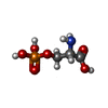 ChemComp-SEP: |
-Macromolecule #6: 3-[(3-CHOLAMIDOPROPYL)DIMETHYLAMMONIO]-1-PROPANESULFONATE
| Macromolecule | Name: 3-[(3-CHOLAMIDOPROPYL)DIMETHYLAMMONIO]-1-PROPANESULFONATE type: ligand / ID: 6 / Number of copies: 1 / Formula: CPS |
|---|---|
| Molecular weight | Theoretical: 614.877 Da |
| Chemical component information | 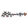 ChemComp-CPS: |
-Macromolecule #7: water
| Macromolecule | Name: water / type: ligand / ID: 7 / Number of copies: 1 / Formula: HOH |
|---|---|
| Molecular weight | Theoretical: 18.015 Da |
| Chemical component information |  ChemComp-HOH: |
-Experimental details
-Structure determination
| Method | cryo EM |
|---|---|
 Processing Processing | single particle reconstruction |
| Aggregation state | particle |
-Sample preparation
| Concentration | 0.1 mg/mL | |||||||||||||||
|---|---|---|---|---|---|---|---|---|---|---|---|---|---|---|---|---|
| Buffer | pH: 7.5 Component:
| |||||||||||||||
| Grid | Model: UltrAuFoil R0./1 / Material: GOLD / Mesh: 300 / Support film - Material: GOLD / Support film - topology: HOLEY / Pretreatment - Type: GLOW DISCHARGE / Pretreatment - Time: 7 sec. / Pretreatment - Atmosphere: OTHER | |||||||||||||||
| Vitrification | Cryogen name: ETHANE / Chamber humidity: 100 % / Chamber temperature: 293 K / Instrument: FEI VITROBOT MARK IV | |||||||||||||||
| Details | Protein is mixed with prepared small unilamellar vesicles |
- Electron microscopy
| Microscope | TFS GLACIOS |
|---|---|
| Specialist optics | Energy filter - Slit width: 20 eV |
| Image recording | Film or detector model: TFS FALCON 4i (4k x 4k) / Number grids imaged: 1 / Number real images: 9197 / Average electron dose: 39.64 e/Å2 |
| Electron beam | Acceleration voltage: 200 kV / Electron source:  FIELD EMISSION GUN FIELD EMISSION GUN |
| Electron optics | Illumination mode: FLOOD BEAM / Imaging mode: BRIGHT FIELD / Cs: 2.7 mm / Nominal defocus max: 2.5 µm / Nominal defocus min: 1.0 µm / Nominal magnification: 130000 |
| Sample stage | Cooling holder cryogen: NITROGEN |
+Image processing

-Atomic model buiding 1
| Refinement | Protocol: AB INITIO MODEL |
|---|---|
| Output model | PDB-36jl: |
