 Movie
Movie Controller

[English] 日本語
 Yorodumi
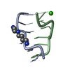
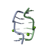
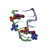
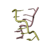
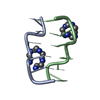
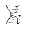
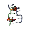
- PDB-362d: THE STRUCTURE OF D(TGCGCA)2 AND A COMPARISON TO OTHER Z-DNA HEXAMERS -
+ Open data

- Basic information
| Entry | Database: PDB / ID: 362d | |||||||||||||||||||||
|---|---|---|---|---|---|---|---|---|---|---|---|---|---|---|---|---|---|---|---|---|---|---|
| Title | THE STRUCTURE OF D(TGCGCA)2 AND A COMPARISON TO OTHER Z-DNA HEXAMERS | |||||||||||||||||||||
 Components Components | DNA (5'-D(* Keywords KeywordsDNA / Z-DNA / DOUBLE HELIX | Function / homology | COBALT HEXAMMINE(III) / DNA |  Function and homology information Function and homology informationMethod |  X-RAY DIFFRACTION / SYNCHROTRON / MOLECULAR REPLACEMENT / Resolution: 1.3 Å X-RAY DIFFRACTION / SYNCHROTRON / MOLECULAR REPLACEMENT / Resolution: 1.3 Å  Authors AuthorsHarper, N.A. / Brannigan, J.A. / Buck, M. / Lewis, R.J. / Moore, M.H. / Schneider, B. |  CitationJournal: Acta Crystallogr.,Sect.D / Year: 1998 CitationJournal: Acta Crystallogr.,Sect.D / Year: 1998Title: Structure of d(TGCGCA)2 and a comparison to other DNA hexamers. Authors: Harper, A. / Brannigan, J.A. / Buck, M. / Hewitt, L. / Lewis, R.J. / Moore, M.H. / Schneider, B. History |
|
- Structure visualization
| Structure viewer | Molecule: MolmilJmol/JSmol |
|---|
- Downloads & links
-Download
| PDBx/mmCIF format | 362d.cif.gz | 26.2 KB | Display | PDBx/mmCIF format |
|---|---|---|---|---|
| PDB format | pdb362d.ent.gz | 19.7 KB | Display | PDB format |
| PDBx/mmJSON format | 362d.json.gz | Tree view | PDBx/mmJSON format | |
| Others |  Other downloads Other downloads |
-Validation report
| Arichive directory | https://data.pdbj.org/pub/pdb/validation_reports/62/362dftp://data.pdbj.org/pub/pdb/validation_reports/62/362d | HTTPS FTP |
|---|
-Related structure data
| Related structure data | 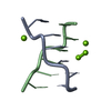 1dcgS S: Starting model for refinement |
|---|---|
| Similar structure data |


-Links
 PDBj
PDBj


- Assembly
| Deposited unit | 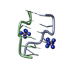
| ||||||||
|---|---|---|---|---|---|---|---|---|---|
| 1 |
| ||||||||
| Unit cell |
|
-Components
| #1: DNA chain | Mass: 1809.218 Da / Num. of mol.: 2 / Source method: obtained synthetically #2: Chemical |   Mass: 161.116 Da / Num. of mol.: 2 / Source method: obtained synthetically / Formula: CoH18N6 Mass: 161.116 Da / Num. of mol.: 2 / Source method: obtained synthetically / Formula: CoH18N6#3: Water | ChemComp-HOH / |  Mass: 18.015 Da / Num. of mol.: 78 / Source method: isolated from a natural source / Formula: H2O Mass: 18.015 Da / Num. of mol.: 78 / Source method: isolated from a natural source / Formula: H2O |
|---|
-Experimental details
-Experiment
| Experiment | Method: X-RAY DIFFRACTION / Number of used crystals: 1 |
|---|
- Sample preparation
| Crystal | Density Matthews: 2 Å3/Da / Density % sol: 38 % | |||||||||||||||||||||||||||||||||||
|---|---|---|---|---|---|---|---|---|---|---|---|---|---|---|---|---|---|---|---|---|---|---|---|---|---|---|---|---|---|---|---|---|---|---|---|---|
| Crystal grow | pH: 6 / Details: pH 6.00 | |||||||||||||||||||||||||||||||||||
| Crystal | *PLUS Density % sol: 38 % | |||||||||||||||||||||||||||||||||||
| Crystal grow | *PLUS Temperature: 291 K / pH: 6 / Method: vapor diffusion, sitting dropDetails: drops of 6 micro litter total volume obtained by combining 3 micro litter of stock solution with 3 micro litter of mother liquor | |||||||||||||||||||||||||||||||||||
| Components of the solutions | *PLUS
|
-Data collection
| Diffraction | Mean temperature: 120 K |
|---|---|
| Diffraction source | Source: SYNCHROTRON / Site: SRS  / Beamline: PX9.6 / Wavelength: 0.87 / Beamline: PX9.6 / Wavelength: 0.87 |
| Detector | Type: MARRESEARCH / Detector: IMAGE PLATE / Date: Sep 1, 1995 |
| Radiation | Monochromatic (M) / Laue (L): M / Scattering type: x-ray |
| Radiation wavelength | Wavelength: 0.87 Å / Relative weight: 1 |
| Reflection | Resolution: 1.3→24 Å / Num. obs: 7024 / % possible obs: 99.2 % / Redundancy: 4 % / Rmerge(I) obs: 0.043 / Net I/σ(I): 20 |
| Reflection shell | Resolution: 1.3→1.32 Å / Redundancy: 4 % / Rmerge(I) obs: 0.078 / Mean I/σ(I) obs: 23.6 / % possible all: 87.4 |
| Reflection shell | *PLUS % possible obs: 87.4 % |
- Processing
| Software |
| |||||||||||||||||||||||||||||||||
|---|---|---|---|---|---|---|---|---|---|---|---|---|---|---|---|---|---|---|---|---|---|---|---|---|---|---|---|---|---|---|---|---|---|---|
| Refinement | Method to determine structure: MOLECULAR REPLACEMENT Starting model: MODIFIED NDB ENTRY ZDF002 (PDB ENTRY 1DCG) Resolution: 1.3→10 Å / Num. parameters: 3018 / Num. restraintsaints: 2782 / σ(F): 0 Details: RESTRAINED ANISOTROPIC REFINEMENT OF NON-HYDROGEN ATOMS. ANISOTROPIC REFINEMENT REDUCED FREE R (NO CUTOFF) BY 1.4%.
| |||||||||||||||||||||||||||||||||
| Solvent computation | Solvent model: MOEWS & KRETSINGER, J.MOL.BIOL.91(1973)201 | |||||||||||||||||||||||||||||||||
| Refine Biso |
| |||||||||||||||||||||||||||||||||
| Refine analyze | Num. disordered residues: 1 / Occupancy sum hydrogen: 138 / Occupancy sum non hydrogen: 332 | |||||||||||||||||||||||||||||||||
| Refinement step | Cycle: LAST / Resolution: 1.3→10 Å
| |||||||||||||||||||||||||||||||||
| Refine LS restraints |
| |||||||||||||||||||||||||||||||||
| Software | *PLUS Name: SHELXL-96 / Classification: refinement | |||||||||||||||||||||||||||||||||
| Refinement | *PLUS Highest resolution: 1.3 Å / Lowest resolution: 10 Å / σ(F): 0 / % reflection Rfree: 10 % / Rfactor obs: 0.12 | |||||||||||||||||||||||||||||||||
| Solvent computation | *PLUS | |||||||||||||||||||||||||||||||||
| Displacement parameters | *PLUS |
