 Movie
Movie Controller

[English] 日本語
 Yorodumi
- PDB-4e4o: Interactions of Ba2+ with a non-self-complementary Z-type DNA duplex -
+ Open data

- Basic information
| Entry | Database: PDB / ID: 4e4o | ||||||
|---|---|---|---|---|---|---|---|
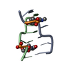
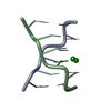
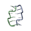
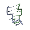
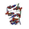
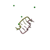
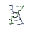
| Title | Interactions of Ba2+ with a non-self-complementary Z-type DNA duplex | ||||||
 Components Components |
| ||||||
 Keywords Keywords | DNA / Z-type DNA double helices / Barium ions | ||||||
| Function / homology | : / DNA Function and homology information Function and homology information | ||||||
| Method |  X-RAY DIFFRACTION / MOLECULAR REPLACEMENT / Resolution: 1.72 Å X-RAY DIFFRACTION / MOLECULAR REPLACEMENT / Resolution: 1.72 Å | ||||||
 Authors Authors | Mandal, P.K. / Venkadesh, S. / Gautham, N. | ||||||
 Citation Citation | Journal: To be Published Title: Interactions of Ba2+ with a non-self-complementary Z-type DNA duplex Authors: Mandal, P.K. / Venkadesh, S. / Gautham, N. | ||||||
| History |
|
- Structure visualization
| Structure viewer | Molecule: MolmilJmol/JSmol |
|---|
- Downloads & links
-Download
| PDBx/mmCIF format | 4e4o.cif.gz | 23.9 KB | Display | PDBx/mmCIF format |
|---|---|---|---|---|
| PDB format | pdb4e4o.ent.gz | 15.7 KB | Display | PDB format |
| PDBx/mmJSON format | 4e4o.json.gz | Tree view | PDBx/mmJSON format | |
| Others |  Other downloads Other downloads |
-Validation report
| Arichive directory | https://data.pdbj.org/pub/pdb/validation_reports/e4/4e4oftp://data.pdbj.org/pub/pdb/validation_reports/e4/4e4o | HTTPS FTP |
|---|
-Related structure data


-Links
 PDBj
PDBj

- Assembly
| Deposited unit | 
| ||||||||
|---|---|---|---|---|---|---|---|---|---|
| 1 | 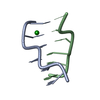
| ||||||||
| 2 | 
| ||||||||
| Unit cell |
|
-Components
| #1: DNA chain | Mass: 1794.206 Da / Num. of mol.: 2 / Source method: obtained synthetically #2: DNA chain | Mass: 1825.216 Da / Num. of mol.: 2 / Source method: obtained synthetically #3: Chemical | ChemComp-BA / |   Mass: 137.327 Da / Num. of mol.: 1 / Source method: obtained synthetically / Formula: Ba Mass: 137.327 Da / Num. of mol.: 1 / Source method: obtained synthetically / Formula: Ba#4: Water | ChemComp-HOH / |  Mass: 18.015 Da / Num. of mol.: 57 / Source method: isolated from a natural source / Formula: H2O Mass: 18.015 Da / Num. of mol.: 57 / Source method: isolated from a natural source / Formula: H2O |
|---|
-Experimental details
-Experiment
| Experiment | Method: X-RAY DIFFRACTION / Number of used crystals: 1 |
|---|
- Sample preparation
| Crystal | Density Matthews: 1.5809 Å3/Da / Density % sol: 22.1971 % |
|---|---|
| Crystal grow | Temperature: 293 K / Method: vapor diffusion, hanging drop / pH: 7 Details: drop: 1mM of duplex DNA, 50mM sodium cacodylate trihydrate buffer (pH 7.0), 12mM barium dichloride dihydrate, 0.5mM spermine, reservoir: 30% 2-methyl-2,4-pentanediol (MPD), VAPOR DIFFUSION, ...Details: drop: 1mM of duplex DNA, 50mM sodium cacodylate trihydrate buffer (pH 7.0), 12mM barium dichloride dihydrate, 0.5mM spermine, reservoir: 30% 2-methyl-2,4-pentanediol (MPD), VAPOR DIFFUSION, HANGING DROP, temperature 293K |
-Data collection
| Diffraction | Mean temperature: 100 K |
|---|---|
| Diffraction source | Source: ROTATING ANODE / Type: BRUKER AXS MICROSTAR / Wavelength: 1.5418 Å |
| Detector | Type: MAR scanner 345 mm plate / Detector: IMAGE PLATE / Date: Oct 6, 2010 / Details: mirror |
| Radiation | Monochromator: graphite / Protocol: SINGLE WAVELENGTH / Monochromatic (M) / Laue (L): M / Scattering type: x-ray |
| Radiation wavelength | Wavelength: 1.5418 Å / Relative weight: 1 |
| Reflection | Resolution: 1.72→25 Å / Num. all: 4897 / Num. obs: 4873 / % possible obs: 99.5 % / Redundancy: 4.71 % / Biso Wilson estimate: 26.9 Å2 / Rmerge(I) obs: 0.0384 / Rsym value: 0.0357 / Net I/σ(I): 9 |
| Reflection shell | Resolution: 1.72→1.78 Å / Redundancy: 4.68 % / Rmerge(I) obs: 0.2076 / Mean I/σ(I) obs: 1.9 / Rsym value: 0.1908 / % possible all: 99.2 |
- Processing
| Software |
| ||||||||||||||||||||||||||||||||||||||||||||||||||||||||||||||||||||||||||||||||||||||||||||||||||||||||||||||||||||||||||||||||||||||||||||||||||||||||||||||||||||||||||
|---|---|---|---|---|---|---|---|---|---|---|---|---|---|---|---|---|---|---|---|---|---|---|---|---|---|---|---|---|---|---|---|---|---|---|---|---|---|---|---|---|---|---|---|---|---|---|---|---|---|---|---|---|---|---|---|---|---|---|---|---|---|---|---|---|---|---|---|---|---|---|---|---|---|---|---|---|---|---|---|---|---|---|---|---|---|---|---|---|---|---|---|---|---|---|---|---|---|---|---|---|---|---|---|---|---|---|---|---|---|---|---|---|---|---|---|---|---|---|---|---|---|---|---|---|---|---|---|---|---|---|---|---|---|---|---|---|---|---|---|---|---|---|---|---|---|---|---|---|---|---|---|---|---|---|---|---|---|---|---|---|---|---|---|---|---|---|---|---|---|---|---|
| Refinement | Method to determine structure: MOLECULAR REPLACEMENT Starting model: fiber model of Z-type DNA built using the program InsightII Resolution: 1.72→23.53 Å / Cor.coef. Fo:Fc: 0.961 / Cor.coef. Fo:Fc free: 0.915 / SU B: 2.645 / SU ML: 0.082 / Cross valid method: THROUGHOUT / ESU R Free: 0.033 / Stereochemistry target values: MAXIMUM LIKELIHOOD
| ||||||||||||||||||||||||||||||||||||||||||||||||||||||||||||||||||||||||||||||||||||||||||||||||||||||||||||||||||||||||||||||||||||||||||||||||||||||||||||||||||||||||||
| Solvent computation | Ion probe radii: 0.8 Å / Shrinkage radii: 0.8 Å / VDW probe radii: 1.4 Å / Solvent model: MASK | ||||||||||||||||||||||||||||||||||||||||||||||||||||||||||||||||||||||||||||||||||||||||||||||||||||||||||||||||||||||||||||||||||||||||||||||||||||||||||||||||||||||||||
| Displacement parameters | Biso mean: 18.7 Å2
| ||||||||||||||||||||||||||||||||||||||||||||||||||||||||||||||||||||||||||||||||||||||||||||||||||||||||||||||||||||||||||||||||||||||||||||||||||||||||||||||||||||||||||
| Refinement step | Cycle: LAST / Resolution: 1.72→23.53 Å
| ||||||||||||||||||||||||||||||||||||||||||||||||||||||||||||||||||||||||||||||||||||||||||||||||||||||||||||||||||||||||||||||||||||||||||||||||||||||||||||||||||||||||||
| Refine LS restraints |
| ||||||||||||||||||||||||||||||||||||||||||||||||||||||||||||||||||||||||||||||||||||||||||||||||||||||||||||||||||||||||||||||||||||||||||||||||||||||||||||||||||||||||||
| LS refinement shell | Resolution: 1.72→1.764 Å / Total num. of bins used: 20
|
