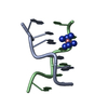
 Movie
Movie Controller


+ Open data

- Basic information
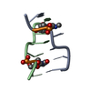
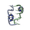
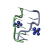
| Entry | Database: PDB / ID: 1omk | ||||||||||||||||||
|---|---|---|---|---|---|---|---|---|---|---|---|---|---|---|---|---|---|---|---|
| Title | The Crystal Structure of d(CACG(5IU)G) | ||||||||||||||||||
 Components Components | 5'-D(* Keywords KeywordsDNA / Z-DNA / 5-IODO-2'-DEOXYURIDINE | Function / homology | COBALT HEXAMMINE(III) / DNA |  Function and homology information Function and homology informationMethod |  X-RAY DIFFRACTION / SYNCHROTRON / MOLECULAR REPLACEMENT / Resolution: 1.3 Å X-RAY DIFFRACTION / SYNCHROTRON / MOLECULAR REPLACEMENT / Resolution: 1.3 Å  Authors AuthorsSchuerman, G. / Van Hecke, K. / Van Meervelt, L. |  CitationJournal: Acta Crystallogr.,Sect.D / Year: 2003 CitationJournal: Acta Crystallogr.,Sect.D / Year: 2003Title: Exploration of the influence of 5-iodo-2'-deoxyuridine incorporation on the structure of d[CACG(IDU)G]. Authors: Schuerman, G. / Van Hecke, K. / Van Meervelt, L. History |
|
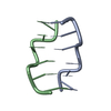
- Structure visualization
| Structure viewer | Molecule: MolmilJmol/JSmol |
|---|
- Downloads & links
-Download
| PDBx/mmCIF format | 1omk.cif.gz | 17.5 KB | Display | PDBx/mmCIF format |
|---|---|---|---|---|
| PDB format | pdb1omk.ent.gz | 10.5 KB | Display | PDB format |
| PDBx/mmJSON format | 1omk.json.gz | Tree view | PDBx/mmJSON format | |
| Others |  Other downloads Other downloads |
-Validation report
| Arichive directory | https://data.pdbj.org/pub/pdb/validation_reports/om/1omkftp://data.pdbj.org/pub/pdb/validation_reports/om/1omk | HTTPS FTP |
|---|
-Related structure data
| Similar structure data |
|---|


-Links
 PDBj
PDBj


- Assembly
| Deposited unit | 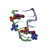
| ||||||||
|---|---|---|---|---|---|---|---|---|---|
| 1 |
| ||||||||
| Unit cell |
|
-Components
| #1: DNA chain | Mass: 1921.087 Da / Num. of mol.: 2 / Source method: obtained synthetically Details: THE OLIGONUCLEOTIDE WAS SYNTHESIZED BY THE REGA-INSTITUTE KULEUVEN #2: Chemical |   Mass: 161.116 Da / Num. of mol.: 2 / Source method: obtained synthetically / Formula: CoH18N6 Mass: 161.116 Da / Num. of mol.: 2 / Source method: obtained synthetically / Formula: CoH18N6#3: Water | ChemComp-HOH / |  Mass: 18.015 Da / Num. of mol.: 43 / Source method: isolated from a natural source / Formula: H2O Mass: 18.015 Da / Num. of mol.: 43 / Source method: isolated from a natural source / Formula: H2O |
|---|
-Experimental details
-Experiment
| Experiment | Method: X-RAY DIFFRACTION / Number of used crystals: 1 |
|---|
- Sample preparation
| Crystal | Density Matthews: 1.49 Å3/Da / Density % sol: 17.43 % | ||||||||||||||||||||||||||||||||||||||||||||||||||||||||||||||||||||||
|---|---|---|---|---|---|---|---|---|---|---|---|---|---|---|---|---|---|---|---|---|---|---|---|---|---|---|---|---|---|---|---|---|---|---|---|---|---|---|---|---|---|---|---|---|---|---|---|---|---|---|---|---|---|---|---|---|---|---|---|---|---|---|---|---|---|---|---|---|---|---|---|
| Crystal grow | Temperature: 290 K / Method: vapor diffusion, hanging drop / pH: 5.5 Details: potassium cacodylate, MPD, magnesium chloride, potassium chloride, cobalt hexamine , pH 5.5, VAPOR DIFFUSION, HANGING DROP, temperature 290.0K | ||||||||||||||||||||||||||||||||||||||||||||||||||||||||||||||||||||||
| Components of the solutions |
| ||||||||||||||||||||||||||||||||||||||||||||||||||||||||||||||||||||||
| Crystal grow | *PLUS pH: 6.9 / Method: vapor diffusion, hanging drop | ||||||||||||||||||||||||||||||||||||||||||||||||||||||||||||||||||||||
| Components of the solutions | *PLUS
|
-Data collection
| Diffraction | Mean temperature: 120 K |
|---|---|
| Diffraction source | Source: SYNCHROTRON / Site: EMBL/DESY, HAMBURG  / Beamline: X11 / Wavelength: 1 Å / Beamline: X11 / Wavelength: 1 Å |
| Detector | Type: MARRESEARCH / Detector: IMAGE PLATE / Date: May 5, 1998 |
| Radiation | Protocol: SINGLE WAVELENGTH / Monochromatic (M) / Laue (L): M / Scattering type: x-ray |
| Radiation wavelength | Wavelength: 1 Å / Relative weight: 1 |
| Reflection | Resolution: 1.3→20 Å / Num. all: 6103 / Num. obs: 5988 / % possible obs: 98.2 % / Redundancy: 22.9 % / Biso Wilson estimate: 22.5 Å2 / Rmerge(I) obs: 0.062 |
| Reflection shell | Resolution: 1.3→1.35 Å / Rmerge(I) obs: 0.165 / Num. unique all: 903 / % possible all: 98.8 |
| Reflection | *PLUS Num. measured all: 137352 |
| Reflection shell | *PLUS % possible obs: 98.8 % |
- Processing
| Software |
| ||||||||||||
|---|---|---|---|---|---|---|---|---|---|---|---|---|---|
| Refinement | Method to determine structure: MOLECULAR REPLACEMENT Starting model: NDB ENTRY ZDFB51 Resolution: 1.3→20 Å / Isotropic thermal model: Isotropic / σ(F): 4 / Details: The iodo atoms are refined anisotropically /
| ||||||||||||
| Refinement step | Cycle: LAST / Resolution: 1.3→20 Å
| ||||||||||||
| Software | *PLUS Name: SHELXL / Version: 93 / Classification: refinement | ||||||||||||
| Refinement | *PLUS Rfactor all: 0.167 | ||||||||||||
| Solvent computation | *PLUS | ||||||||||||
| Displacement parameters | *PLUS |
