 Movie
Movie Controller

[English] 日本語
 Yorodumi
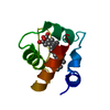
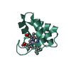
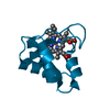
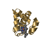
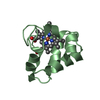
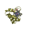
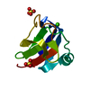
- PDB-351c: STRUCTURE OF CYTOCHROME C551 FROM P. AERUGINOSA REFINED AT 1.6 AN... -
+ Open data

- Basic information
| Entry | Database: PDB / ID: 351c | |||||||||
|---|---|---|---|---|---|---|---|---|---|---|
| Title | STRUCTURE OF CYTOCHROME C551 FROM P. AERUGINOSA REFINED AT 1.6 ANGSTROMS RESOLUTION AND COMPARISON OF THE TWO REDOX FORMS | |||||||||
 Components Components | CYTOCHROME C551 | |||||||||
 Keywords Keywords | ELECTRON TRANSPORT | |||||||||
| Function / homology |  Function and homology information Function and homology informationelectron transfer activity / periplasmic space / iron ion binding / heme binding Similarity search - Function | |||||||||
| Biological species |   Pseudomonas aeruginosa (bacteria) Pseudomonas aeruginosa (bacteria) | |||||||||
| Method |  X-RAY DIFFRACTION / Resolution: 1.6 Å X-RAY DIFFRACTION / Resolution: 1.6 Å | |||||||||
 Authors Authors | Matsuura, Y. / Takano, T. / Dickerson, R.E. | |||||||||
 Citation Citation | Journal: J.Mol.Biol. / Year: 1982 Title: Structure of cytochrome c551 from Pseudomonas aeruginosa refined at 1.6 A resolution and comparison of the two redox forms. Authors: Matsuura, Y. / Takano, T. / Dickerson, R.E. #1: Journal: Proc.Natl.Acad.Sci.USA / Year: 1978Title: Pseudomonas Cytochrome C551 at 2.0 Angstroms Resolution. Enlargement of the Cytochrome C Family Authors: Almassy, R.J. / Dickerson, R.E. #2: Journal: J.Mol.Biol. / Year: 1976Title: The Cytochrome Fold and the Evolution of Bacterial Energy Metabolism Authors: Dickerson, R.E. / Timkovich, R. / Almassy, R.J. | |||||||||
| History |
|
- Structure visualization
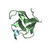
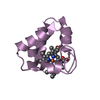
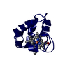
| Structure viewer | Molecule: MolmilJmol/JSmol |
|---|
- Downloads & links
-Download
| PDBx/mmCIF format | 351c.cif.gz | 30.6 KB | Display | PDBx/mmCIF format |
|---|---|---|---|---|
| PDB format | pdb351c.ent.gz | 19.6 KB | Display | PDB format |
| PDBx/mmJSON format | 351c.json.gz | Tree view | PDBx/mmJSON format | |
| Others |  Other downloads Other downloads |
-Validation report
| Arichive directory | https://data.pdbj.org/pub/pdb/validation_reports/51/351cftp://data.pdbj.org/pub/pdb/validation_reports/51/351c | HTTPS FTP |
|---|
-Related structure data

-Links
 PDBj
PDBj


- Assembly
| Deposited unit | 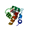
| ||||||||
|---|---|---|---|---|---|---|---|---|---|
| 1 |
| ||||||||
| Unit cell |
|
-Components
| #1: Protein | Mass: 8704.892 Da / Num. of mol.: 1 Source method: isolated from a genetically manipulated source Source: (gene. exp.) Pseudomonas aeruginosa (bacteria) / References: UniProt: P00099 | ||||
|---|---|---|---|---|---|
| #2: Chemical |   Mass: 616.487 Da / Num. of mol.: 1 / Source method: obtained synthetically / Formula: C34H32FeN4O4 Mass: 616.487 Da / Num. of mol.: 1 / Source method: obtained synthetically / Formula: C34H32FeN4O4#3: Water | ChemComp-HOH / |  Mass: 18.015 Da / Num. of mol.: 67 / Source method: isolated from a natural source / Formula: H2O Mass: 18.015 Da / Num. of mol.: 67 / Source method: isolated from a natural source / Formula: H2OHas protein modification | Y | |
-Experimental details
-Experiment
| Experiment | Method: X-RAY DIFFRACTION |
|---|
- Sample preparation
| Crystal | Density Matthews: 2.07 Å3/Da / Density % sol: 40.6 % | ||||||||||||||||||||||||
|---|---|---|---|---|---|---|---|---|---|---|---|---|---|---|---|---|---|---|---|---|---|---|---|---|---|
| Crystal grow | *PLUS Method: microdialysis / PH range low: 5.7 / PH range high: 5.6 | ||||||||||||||||||||||||
| Components of the solutions | *PLUS
|
-Data collection
| Reflection | *PLUS Num. obs: 8670 |
|---|
- Processing
| Refinement | Highest resolution: 1.6 Å /
| ||||||||||||
|---|---|---|---|---|---|---|---|---|---|---|---|---|---|
| Refinement step | Cycle: LAST / Highest resolution: 1.6 Å
| ||||||||||||
| Refinement | *PLUS Rfactor obs: 0.195 | ||||||||||||
| Solvent computation | *PLUS | ||||||||||||
| Displacement parameters | *PLUS |
