 Movie
Movie Controller

[English] 日本語
 Yorodumi
- PDB-3zow: Crystal Structure of Wild Type Nitrosomonas europaea Cytochrome c552 -
+ Open data

- Basic information
| Entry | Database: PDB / ID: 3zow | ||||||
|---|---|---|---|---|---|---|---|
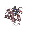
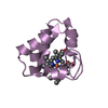
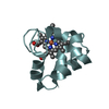
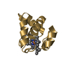
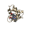
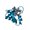
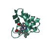
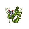
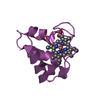
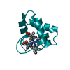
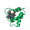
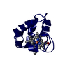
| Title | Crystal Structure of Wild Type Nitrosomonas europaea Cytochrome c552 | ||||||
 Components Components | CYTOCHROME C-552 | ||||||
 Keywords Keywords | ELECTRON TRANSPORT / HEMEPROTEIN | ||||||
| Function / homology |  Function and homology information Function and homology informationelectron transfer activity / periplasmic space / iron ion binding / heme binding Similarity search - Function | ||||||
| Biological species |  NITROSOMONAS EUROPAEA (bacteria) NITROSOMONAS EUROPAEA (bacteria) | ||||||
| Method |  X-RAY DIFFRACTION / SYNCHROTRON / MOLECULAR REPLACEMENT / Resolution: 2.35 Å X-RAY DIFFRACTION / SYNCHROTRON / MOLECULAR REPLACEMENT / Resolution: 2.35 Å | ||||||
 Authors Authors | Hersleth, H.-P. / Can, M. / Krucinska, J. / Zoppellaro, G. / Andersen, N.H. / Karlsen, S. / Wedekind, J.E. / Andersson, K.K. / Bren, K.L. | ||||||
 Citation Citation | Journal: Chembiochem / Year: 2013 Title: Structural Characterization of Nitrosomonas Europaea Cytochrome C-552 Variants with Marked Differences in Electronic Structure. Authors: Can, M. / Krucinska, J. / Zoppellaro, G. / Andersen, N.H. / Wedekind, J.E. / Hersleth, H.-P. / Andersson, K.K. / Bren, K.L. | ||||||
| History |
|
- Structure visualization
| Structure viewer | Molecule: MolmilJmol/JSmol |
|---|
- Downloads & links
-Download
| PDBx/mmCIF format | 3zow.cif.gz | 548.7 KB | Display | PDBx/mmCIF format |
|---|---|---|---|---|
| PDB format | pdb3zow.ent.gz | 465.6 KB | Display | PDB format |
| PDBx/mmJSON format | 3zow.json.gz | Tree view | PDBx/mmJSON format | |
| Others |  Other downloads Other downloads |
-Validation report
| Arichive directory | https://data.pdbj.org/pub/pdb/validation_reports/zo/3zowftp://data.pdbj.org/pub/pdb/validation_reports/zo/3zow | HTTPS FTP |
|---|
-Related structure data
| Related structure data |  3zoxSC  3zoyC  4jcgC S: Starting model for refinement C: citing same article ( |
|---|---|
| Similar structure data |


-Links
 PDBj
PDBj


- Assembly


-Components
| #1: Protein | Mass: 8491.702 Da / Num. of mol.: 18 / Source method: isolated from a natural source / Source: (natural) NITROSOMONAS EUROPAEA (bacteria)References: UniProt: P95339, nitrite reductase (cytochrome; ammonia-forming) #2: Chemical | ChemComp-HEC /   Mass: 618.503 Da / Num. of mol.: 18 / Source method: obtained synthetically / Formula: C34H34FeN4O4 Mass: 618.503 Da / Num. of mol.: 18 / Source method: obtained synthetically / Formula: C34H34FeN4O4#3: Water | ChemComp-HOH / |  Mass: 18.015 Da / Num. of mol.: 216 / Source method: isolated from a natural source / Formula: H2O Mass: 18.015 Da / Num. of mol.: 216 / Source method: isolated from a natural source / Formula: H2OHas protein modification | Y | |
|---|
-Experimental details
-Experiment
| Experiment | Method: X-RAY DIFFRACTION / Number of used crystals: 1 |
|---|
- Sample preparation
| Crystal | Density Matthews: 2.57 Å3/Da / Density % sol: 51.9 % / Description: NONE |
|---|---|
| Crystal grow | pH: 8 / Details: 3 M AMMONIUM SULFATE, 10 MM TRIS-HCL PH 8 |
-Data collection
| Diffraction | Mean temperature: 100 K |
|---|---|
| Diffraction source | Source: SYNCHROTRON / Site: EMBL/DESY, HAMBURG  / Beamline: X11 / Wavelength: 0.8496 / Beamline: X11 / Wavelength: 0.8496 |
| Detector | Type: MARRESEARCH / Detector: CCD / Date: Oct 24, 2001 |
| Radiation | Protocol: SINGLE WAVELENGTH / Monochromatic (M) / Laue (L): M / Scattering type: x-ray |
| Radiation wavelength | Wavelength: 0.8496 Å / Relative weight: 1 |
| Reflection | Resolution: 2.35→38.8 Å / Num. obs: 68939 / % possible obs: 97.1 % / Observed criterion σ(I): 6 / Redundancy: 3.3 % / Rmerge(I) obs: 0.06 / Net I/σ(I): 13.6 |
| Reflection shell | Resolution: 2.35→2.48 Å / Redundancy: 2.8 % / Rmerge(I) obs: 0.3 / Mean I/σ(I) obs: 3.2 / % possible all: 89.1 |
- Processing
| Software |
| ||||||||||||||||||||||||||||||||||||||||||||||||||||||||||||||||||||||||||||||||||||||||||||||||||||||||||||||||||||||||||||||||||||||||||||||||||||||||||||||||||||||||||||||||||||||
|---|---|---|---|---|---|---|---|---|---|---|---|---|---|---|---|---|---|---|---|---|---|---|---|---|---|---|---|---|---|---|---|---|---|---|---|---|---|---|---|---|---|---|---|---|---|---|---|---|---|---|---|---|---|---|---|---|---|---|---|---|---|---|---|---|---|---|---|---|---|---|---|---|---|---|---|---|---|---|---|---|---|---|---|---|---|---|---|---|---|---|---|---|---|---|---|---|---|---|---|---|---|---|---|---|---|---|---|---|---|---|---|---|---|---|---|---|---|---|---|---|---|---|---|---|---|---|---|---|---|---|---|---|---|---|---|---|---|---|---|---|---|---|---|---|---|---|---|---|---|---|---|---|---|---|---|---|---|---|---|---|---|---|---|---|---|---|---|---|---|---|---|---|---|---|---|---|---|---|---|---|---|---|---|
| Refinement | Method to determine structure: MOLECULAR REPLACEMENT Starting model: PDB ENTRY 3ZOX Resolution: 2.35→35.22 Å / Cor.coef. Fo:Fc: 0.932 / Cor.coef. Fo:Fc free: 0.891 / SU B: 13.213 / SU ML: 0.188 / Cross valid method: THROUGHOUT / ESU R: 0.382 / ESU R Free: 0.263 / Stereochemistry target values: MAXIMUM LIKELIHOOD Details: HYDROGENS HAVE BEEN ADDED IN THE RIDING POSITIONS. HYDROGENS HAVE BEEN USED IF PRESENT IN THE INPUT. IT IS COMMON IN PUBLICATIONS ETC. TO START THE NUMBERING OF THE CYTOCHROME C552 ...Details: HYDROGENS HAVE BEEN ADDED IN THE RIDING POSITIONS. HYDROGENS HAVE BEEN USED IF PRESENT IN THE INPUT. IT IS COMMON IN PUBLICATIONS ETC. TO START THE NUMBERING OF THE CYTOCHROME C552 NITROSOMONAS EUROPAEA WITH THE FIRST RESIDUE IN THE SEQUENCE BEING NUMBERED AS RESIDUE 3. SINGLE-CRYSTAL UV-VIS SPECTRA HAVE BEEN RECORDED BEFORE AND AFTER EXPOSURE TO X-RAYS SHOWING THE RADIATION-INFLUENCE OF THE FERRIC CYTOCHROME C552 CRYSTALS SEE JRNL.
| ||||||||||||||||||||||||||||||||||||||||||||||||||||||||||||||||||||||||||||||||||||||||||||||||||||||||||||||||||||||||||||||||||||||||||||||||||||||||||||||||||||||||||||||||||||||
| Solvent computation | Ion probe radii: 0.8 Å / Shrinkage radii: 0.8 Å / VDW probe radii: 1.2 Å / Solvent model: MASK | ||||||||||||||||||||||||||||||||||||||||||||||||||||||||||||||||||||||||||||||||||||||||||||||||||||||||||||||||||||||||||||||||||||||||||||||||||||||||||||||||||||||||||||||||||||||
| Displacement parameters | Biso mean: 37.493 Å2
| ||||||||||||||||||||||||||||||||||||||||||||||||||||||||||||||||||||||||||||||||||||||||||||||||||||||||||||||||||||||||||||||||||||||||||||||||||||||||||||||||||||||||||||||||||||||
| Refinement step | Cycle: LAST / Resolution: 2.35→35.22 Å
| ||||||||||||||||||||||||||||||||||||||||||||||||||||||||||||||||||||||||||||||||||||||||||||||||||||||||||||||||||||||||||||||||||||||||||||||||||||||||||||||||||||||||||||||||||||||
| Refine LS restraints |
|