 Movie
Movie Controller

[English] 日本語
 Yorodumi
- PDB-2zz3: Covalent complex of orotidine monophosphate decarboxylase D70A mu... -
+ Open data

- Basic information
| Entry | Database: PDB / ID: 2zz3 | ||||||
|---|---|---|---|---|---|---|---|
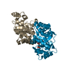
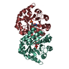
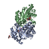
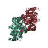
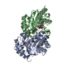
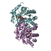
| Title | Covalent complex of orotidine monophosphate decarboxylase D70A mutant from M. thermoautotrophicus with 6-cyano-UMP | ||||||
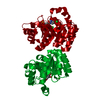
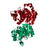
 Components Components | Orotidine 5'-phosphate decarboxylase | ||||||
 Keywords Keywords | LYASE / ODCase / OMPDCase / OMPDC / Decarboxylase / Pyrimidine biosynthesis | ||||||
| Function / homology |  Function and homology information Function and homology informationorotidine-5'-phosphate decarboxylase / orotidine-5'-phosphate decarboxylase activity / 'de novo' UMP biosynthetic process / 'de novo' pyrimidine nucleobase biosynthetic process / cytosol Similarity search - Function | ||||||
| Biological species |   Methanothermobacter thermautotrophicus (archaea) Methanothermobacter thermautotrophicus (archaea) | ||||||
| Method |  X-RAY DIFFRACTION / SYNCHROTRON / FOURIER SYNTHESIS / Resolution: 1.8 Å X-RAY DIFFRACTION / SYNCHROTRON / FOURIER SYNTHESIS / Resolution: 1.8 Å | ||||||
 Authors Authors | Fujihashi, M. / Pai, E.F. | ||||||
 Citation Citation | Journal: J.Mol.Biol. / Year: 2009 Title: Structural characterization of the molecular events during a slow substrate-product transition in orotidine 5'-monophosphate decarboxylase Authors: Fujihashi, M. / Wei, L. / Kotra, L.P. / Pai, E.F. | ||||||
| History |
|
- Structure visualization
| Structure viewer | Molecule: MolmilJmol/JSmol |
|---|
- Downloads & links
-Download
| PDBx/mmCIF format | 2zz3.cif.gz | 104.7 KB | Display | PDBx/mmCIF format |
|---|---|---|---|---|
| PDB format | pdb2zz3.ent.gz | 79.7 KB | Display | PDB format |
| PDBx/mmJSON format | 2zz3.json.gz | Tree view | PDBx/mmJSON format | |
| Others |  Other downloads Other downloads |
-Validation report
| Arichive directory | https://data.pdbj.org/pub/pdb/validation_reports/zz/2zz3ftp://data.pdbj.org/pub/pdb/validation_reports/zz/2zz3 | HTTPS FTP |
|---|
-Related structure data
| Related structure data |  2zz1C 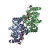 2zz2C 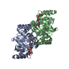 2zz4C 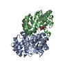 2zz5C 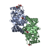 2zz6C  2zz7C 1x1zS S: Starting model for refinement C: citing same article ( |
|---|---|
| Similar structure data |


-Links
 PDBj
PDBj
- Assembly
| Deposited unit | 
| ||||||||
|---|---|---|---|---|---|---|---|---|---|
| 1 |
| ||||||||
| Unit cell |
|
-Components
| #1: Protein | Mass: 27335.391 Da / Num. of mol.: 2 / Mutation: D70A,R226L,I227N Source method: isolated from a genetically manipulated source Source: (gene. exp.) Methanothermobacter thermautotrophicus (archaea)Plasmid: pET15b / Production host:  References: UniProt: O26232, orotidine-5'-phosphate decarboxylase #2: Chemical | 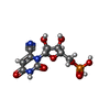  Mass: 349.191 Da / Num. of mol.: 2 / Source method: obtained synthetically / Formula: C10H12N3O9P Mass: 349.191 Da / Num. of mol.: 2 / Source method: obtained synthetically / Formula: C10H12N3O9P#3: Chemical | ChemComp-GOL /   Mass: 92.094 Da / Num. of mol.: 4 / Source method: obtained synthetically / Formula: C3H8O3 Mass: 92.094 Da / Num. of mol.: 4 / Source method: obtained synthetically / Formula: C3H8O3#4: Water | ChemComp-HOH / |  Mass: 18.015 Da / Num. of mol.: 239 / Source method: isolated from a natural source / Formula: H2O Mass: 18.015 Da / Num. of mol.: 239 / Source method: isolated from a natural source / Formula: H2OHas protein modification | Y | Nonpolymer details | THIS COMPLEX WAS PREPARED BY MIXING D70A MUTANT ENZYME AND 6-CYANO-UMP. THE CN GROUP WAS REPLACED ...THIS COMPLEX WAS PREPARED BY MIXING D70A MUTANT ENZYME AND 6-CYANO-UMP. THE CN GROUP WAS REPLACED BY THE AMINO GROUP OF LYS 72. | Sequence details | ACCORDING TO DEPOSITORS, PRO101 IS CORRECT AND UNIPORT IS PROBABLY INCORRECT AT THIS POSITION. ...ACCORDING TO DEPOSITORS | |
|---|
-Experimental details
-Experiment
| Experiment | Method: X-RAY DIFFRACTION / Number of used crystals: 1 |
|---|
- Sample preparation
| Crystal | Density Matthews: 2.02 Å3/Da / Density % sol: 39.08 % |
|---|---|
| Crystal grow | Temperature: 298 K / Method: vapor diffusion Details: 1.2-1.36M Sodium Citrate, 5% dioxiane, pH 6.0-8.5, VAPOR DIFFUSION, temperature 298K PH range: 6.0-8.5 |
-Data collection
| Diffraction source | Source: SYNCHROTRON / Site: APS  / Beamline: 14-ID-B / Wavelength: 1.127 Å / Beamline: 14-ID-B / Wavelength: 1.127 Å |
|---|---|
| Detector | Type: MAR CCD 165 mm / Detector: CCD / Date: Mar 7, 2004 / Details: default |
| Radiation | Protocol: SINGLE WAVELENGTH / Monochromatic (M) / Laue (L): M / Scattering type: x-ray |
| Radiation wavelength | Wavelength: 1.127 Å / Relative weight: 1 |
| Reflection | Resolution: 1.8→100 Å / Num. obs: 38640 / % possible obs: 87.9 % / Redundancy: 3.62 % / Biso Wilson estimate: 13.6 Å2 / Rsym value: 0.063 / Net I/σ(I): 18.3 |
| Reflection shell | Resolution: 1.8→1.83 Å / Mean I/σ(I) obs: 3 / Rsym value: 0.273 / % possible all: 57 |
- Processing
| Software |
| ||||||||||||||||||||||||||||||||||||||||||||||||||||||||||||||||||||||||||||||||
|---|---|---|---|---|---|---|---|---|---|---|---|---|---|---|---|---|---|---|---|---|---|---|---|---|---|---|---|---|---|---|---|---|---|---|---|---|---|---|---|---|---|---|---|---|---|---|---|---|---|---|---|---|---|---|---|---|---|---|---|---|---|---|---|---|---|---|---|---|---|---|---|---|---|---|---|---|---|---|---|---|---|
| Refinement | Method to determine structure: FOURIER SYNTHESIS Starting model: 1x1z Resolution: 1.8→22.19 Å / Rfactor Rfree error: 0.004 / Data cutoff high absF: 1322201.97 / Data cutoff low absF: 0 / Isotropic thermal model: RESTRAINED / Cross valid method: THROUGHOUT / σ(F): 0 / Stereochemistry target values: Engh & Huber
| ||||||||||||||||||||||||||||||||||||||||||||||||||||||||||||||||||||||||||||||||
| Solvent computation | Solvent model: FLAT MODEL / Bsol: 47.1215 Å2 / ksol: 0.419454 e/Å3 | ||||||||||||||||||||||||||||||||||||||||||||||||||||||||||||||||||||||||||||||||
| Displacement parameters | Biso mean: 16.6 Å2
| ||||||||||||||||||||||||||||||||||||||||||||||||||||||||||||||||||||||||||||||||
| Refine analyze |
| ||||||||||||||||||||||||||||||||||||||||||||||||||||||||||||||||||||||||||||||||
| Refinement step | Cycle: LAST / Resolution: 1.8→22.19 Å
| ||||||||||||||||||||||||||||||||||||||||||||||||||||||||||||||||||||||||||||||||
| Refine LS restraints |
| ||||||||||||||||||||||||||||||||||||||||||||||||||||||||||||||||||||||||||||||||
| LS refinement shell | Resolution: 1.8→1.86 Å / Rfactor Rfree error: 0.024 / Total num. of bins used: 10
| ||||||||||||||||||||||||||||||||||||||||||||||||||||||||||||||||||||||||||||||||
| Xplor file |
|
