| Software | | Name | Version | Classification |
|---|
| SOLVE | | phasing| CNS | 0.5 | refinement| DENZO | | data reduction| SCALEPACK | | data scaling | | | |
|
|---|
| Refinement | Resolution: 1.8→30 Å / Rfactor Rfree error: 0.005 / Data cutoff high absF: 914445.28 / Data cutoff low absF: 0 / Isotropic thermal model: RESTRAINED / Cross valid method: THROUGHOUT / σ(F): 0 / σ(I): 0 / Stereochemistry target values: CNS_0.5 library
| Rfactor | Num. reflection | % reflection | Selection details |
|---|
| Rfree | 0.222 | 1980 | 10.1 % | RANDOM |
|---|
| Rwork | 0.2 | - | - | - |
|---|
| all | 0.2 | 19510 | - | - |
|---|
| obs | 0.2 | 19510 | 99.1 % | - |
|---|
|
|---|
| Solvent computation | Solvent model: FLAT MODEL / Bsol: 43.05 Å2 / ksol: 0.3788 e/Å3 |
|---|
| Displacement parameters | Biso mean: 21 Å2
| Baniso -1 | Baniso -2 | Baniso -3 |
|---|
| 1- | -0.63 Å2 | 0 Å2 | 0 Å2 |
|---|
| 2- | - | -0.44 Å2 | 0 Å2 |
|---|
| 3- | - | - | 1.07 Å2 |
|---|
|
|---|
| Refine analyze | | Free | Obs |
|---|
| Luzzati coordinate error | 0.22 Å | 0.19 Å |
|---|
| Luzzati d res low | - | 5 Å |
|---|
| Luzzati sigma a | 0.07 Å | -0.03 Å |
|---|
|
|---|
| Refinement step | Cycle: LAST / Resolution: 1.8→30 Å
| Protein | Nucleic acid | Ligand | Solvent | Total |
|---|
| Num. atoms | 1624 | 0 | 0 | 217 | 1841 |
|---|
|
|---|
| Refine LS restraints | | Refine-ID | Type | Dev ideal | Dev ideal target |
|---|
| X-RAY DIFFRACTION | c_bond_d| 0.012 | | | X-RAY DIFFRACTION | c_angle_deg| 1.5 | | | X-RAY DIFFRACTION | c_dihedral_angle_d| 23 | | | X-RAY DIFFRACTION | c_improper_angle_d| 1.1 | | | X-RAY DIFFRACTION | c_mcbond_it| 1.2 | 1.5 | | X-RAY DIFFRACTION | c_mcangle_it| 1.85 | 2 | | X-RAY DIFFRACTION | c_scbond_it| 2.23 | 2 | | X-RAY DIFFRACTION | c_scangle_it| 3.26 | 2.5 | | | | | | | | |
|
|---|
| LS refinement shell | Resolution: 1.8→1.91 Å / Rfactor Rfree error: 0.013 / Total num. of bins used: 6
| Rfactor | Num. reflection | % reflection |
|---|
| Rfree | 0.239 | 325 | 10.2 % |
|---|
| Rwork | 0.198 | 2869 | - |
|---|
| obs | - | - | 99.8 % |
|---|
|
|---|
| Xplor file | | Refine-ID | Serial no | Param file | Topol file |
|---|
| X-RAY DIFFRACTION | 1 | PROTEIN_REP.PARAMPROTEIN.TOP| X-RAY DIFFRACTION | 2 | WATER_REP.PARAM| WATER.TOP | | | |
|
|---|
| Software | *PLUS Name: CNS / Version: 0.5 / Classification: refinement |
|---|
| Refinement | *PLUS σ(F): 0 / % reflection Rfree: 10.1 % / Rfactor obs: 0.2 / Rfactor Rwork: 0.2 |
|---|
| Solvent computation | *PLUS |
|---|
| Displacement parameters | *PLUS Biso mean: 21 Å2 |
|---|
| Refine LS restraints | *PLUS | Refine-ID | Type | Dev ideal | Dev ideal target |
|---|
| X-RAY DIFFRACTION | c_bond_d| 0.012 | | | X-RAY DIFFRACTION | c_angle_deg| 1.5 | | | X-RAY DIFFRACTION | c_dihedral_angle_d | | | X-RAY DIFFRACTION | c_mcbond_it | 1.5 | | X-RAY DIFFRACTION | c_scbond_it | 2 | | X-RAY DIFFRACTION | c_mcangle_it | 2 | | X-RAY DIFFRACTION | c_scangle_it | 2.5 | | X-RAY DIFFRACTION | c_dihedral_angle_deg| 23 | | | X-RAY DIFFRACTION | c_improper_angle_d | | | X-RAY DIFFRACTION | c_improper_angle_deg| 1.1 | | | | | | | | | | | |
|
|---|
| LS refinement shell | *PLUS Rfactor Rfree: 0.239 / % reflection Rfree: 10.2 % / Rfactor Rwork: 0.198 |
|---|
 Movie
Movie Controller


 Open data
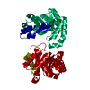
 Basic information
 Components
 Keywords
 Function and homology information
 Methanothermobacter thermautotrophicus (archaea)
 X-RAY DIFFRACTION /
  Authors
 Citation
 Structure visualization
 Downloads & links
 Other downloads


 PDBj
PDBj
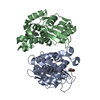
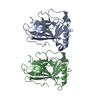
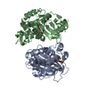
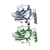
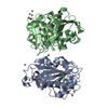
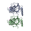
 Assembly
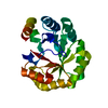


 Mass: 18.015 Da / Num. of mol.: 217 / Source method: isolated from a natural source / Formula: H2O
 Sample preparation
 / Type:
  Processing