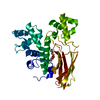
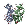
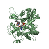
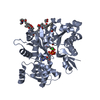
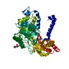
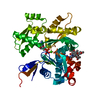
F-ACTIN IS A HELICAL POLYMER OF ACTIN MOLECULES. THE Z-AXIS OF THE ACTIN COORDINATES SHOWN IN THIS PDB-FILE IS A HELIX AXIS. THE NEXT SUBUNIT IN F-ACTIN IS POSITIONED BY 166.4 DEGREE RIGHT ROTATION ABOUT THE HELIX AXIS AND 27.59 ANGSTROM SHIFT ALONG THE HELIX AXIS.
-
Components
#1: Protein
Actin, alphaskeletalmuscle / Alpha-actin-1
Mass: 41875.633 Da / Num. of mol.: 1 / Source method: isolated from a natural source / Details: Rabbit skeletal muscle / Source: (natural) Oryctolagus cuniculus (rabbit) / References: UniProt: P68135
Resolution: 3.3→56 Å / Rfactor all: 0.143 Details: THE DIFFRACTION DATA WERE EXTRACTED USING THE PROPER HELICAL SYMMETRY SUCH AS A SELECTION RULE L=-153N+331M AND A REPEAT DISTANCE 9135 ANGSTRUM. THE LAYER-LINE DISTRIBUTION BY THE SELECTOIN ...Details: THE DIFFRACTION DATA WERE EXTRACTED USING THE PROPER HELICAL SYMMETRY SUCH AS A SELECTION RULE L=-153N+331M AND A REPEAT DISTANCE 9135 ANGSTRUM. THE LAYER-LINE DISTRIBUTION BY THE SELECTOIN RULE IS CLOSE TO THAT BY L=-6N+13M IN PRINCILE ONE LAYER-LINE BY L=-6N+13M SEPARATES SEVERAL LAYER-LINES BY L=-153N+331M. BUT THE SEPARATIONS WERE NOT CLEAR AND THE LAYER-LINE INTENSITYIES WERE EXTRACTED AS "GROUPING LAYER-LINE INTENSITIES" FROM EXPERIMANTAL PATTERNS. THEN THE LAYER-LINE INTENSTITIES WERE HANDLED AS "GROUPING LAYER-LINE INTENSITIES" RE-INDEXED BY L=-6N+13M. THE INITIAL MODELS OF F-ACTIN WERE MADE FROM THE THREE CRYSTAL STRUCTURE OF 1J6Z, 2BTF AND 1HLU. FIRST THE RADIUS AND THREE ORIENTAIONS OF THE SUBUNIT IN F-ACTIN WERE DETERMINED BY THE RIGID BODY SEARCH USING THE DIFFRACTION DATA OF 56-7.2 ANGSTROM. NEXT THE SUBUNIT COMFORMATIONS WERE SEARCHED ALONG THE ADDITIONAL 12 LOWEST ELASTIC NORMAL MODES OF ACTIN MONOMER VIBRATIONS BY COMPARISON BETWEEN THE EXPERIMETAL DIFFRACTION DATA AND THE CALCULATION AT 56-7.2 ANGSTROM. THE DIFFRACTION WAS CALCULATED USING A REPEAT DISTANCE OF 9135A AND A SELECTION RULE L=-153N+331M. THE CALCULATED LAYER-LINE INTENSTITIES ALSO WERE HANDLED A "GROUPING LAYER-LINE INTENSITIES" RE-INDEXED BY L=- 6N+13M. WE CAN NOT FIND THE GOOD SOLVENT MODEL AROUND F-ACTIN. THEN WE USED TWO KINDS OF ATOMIC SCATTERING FACTORS AT THE HIGH RESOLUTION AND THE LOW RESOLUTION DATA.THE MOLECULAR DYNAMICS REFINEMENT AND ENERGY-MINIMIZATION WERE REPEATED IN TRUN USING THE LOW RESOLUTION DATA (RADIALLY 6.5-56 A FROM ORIGIN) AND THE HIGH RESOLUTION DATA (RADIALLY 3.3-5.5 A AND LATERALLY FROM THE MERIDIAN TO 5.5 A) BY FX-PLOR. TO JUDGE THE MODELS, TWO KINDS OF R-FACTORS WERE CALUCLATED AGAINT THE DIFFRACTION PATTERN TO AVOID THE DECOMVOLUTION ERROR. R-FACTOR-FIT IS AN R-FACTOR AGAINST FITTING AREA (THE AREA RADIAL 6.5-56 and 3.6-5.5A AND LATERALLY FROM THE MERIDIAN TO 5.8 A). THE R-FACTOR-NON-FIT IS AN R-FACTOR AGAINST NON-FITTING AREA (THE AREA RADIALLY 5.5 - 6.5 A AND LATERALLY TO 15 A). R-FACTOR-FIT : 0.143 R-FACTOR-NON-FIT : 0.207
Refinement step
Cycle: LAST / Resolution: 3.3→56 Å
Protein
Nucleic acid
Ligand
Solvent
Total
Num. atoms
2933
0
28
0
2961
+
About Yorodumi
-
News
-
Feb 9, 2022. New format data for meta-information of EMDB entries
New format data for meta-information of EMDB entries
Version 3 of the EMDB header file is now the official format.
The previous official version 1.9 will be removed from the archive.
In the structure databanks used in Yorodumi, some data are registered as the other names, "COVID-19 virus" and "2019-nCoV". Here are the details of the virus and the list of structure data.
Jan 31, 2019. EMDB accession codes are about to change! (news from PDBe EMDB page)
EMDB accession codes are about to change! (news from PDBe EMDB page)
The allocation of 4 digits for EMDB accession codes will soon come to an end. Whilst these codes will remain in use, new EMDB accession codes will include an additional digit and will expand incrementally as the available range of codes is exhausted. The current 4-digit format prefixed with “EMD-” (i.e. EMD-XXXX) will advance to a 5-digit format (i.e. EMD-XXXXX), and so on. It is currently estimated that the 4-digit codes will be depleted around Spring 2019, at which point the 5-digit format will come into force.
The EM Navigator/Yorodumi systems omit the EMD- prefix.
Related info.:Q: What is EMD? / ID/Accession-code notation in Yorodumi/EM Navigator
Yorodumi is a browser for structure data from EMDB, PDB, SASBDB, etc.
This page is also the successor to EM Navigator detail page, and also detail information page/front-end page for Omokage search.
The word "yorodu" (or yorozu) is an old Japanese word meaning "ten thousand". "mi" (miru) is to see.
Related info.:EMDB / PDB / SASBDB / Comparison of 3 databanks / Yorodumi Search / Aug 31, 2016. New EM Navigator & Yorodumi / Yorodumi Papers / Jmol/JSmol / Function and homology information / Changes in new EM Navigator and Yorodumi
 Movie
Movie Controller


 Open data
 Basic information
 Components
 Keywords
 Function and homology information
 FIBER DIFFRACTION /
  Authors
 Citation
 Structure visualization
 Downloads & links
 Other downloads
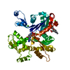


 PDBj
PDBj


 Assembly

 Mass: 427.201 Da / Num. of mol.: 1 / Source method: obtained synthetically / Formula: C10H15N5O10P2 / Comment: ADP, energy-carrying molecule*YM
 Mass: 40.078 Da / Num. of mol.: 1 / Source method: obtained synthetically / Formula: Ca
 Processing