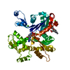
toxin sequestering activity / sporulation resulting in formation of a cellular spore / plasma membrane Similarity search - Function
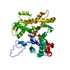
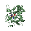
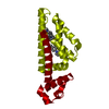
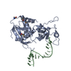
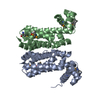
Alpha-Beta Plaits - #2720 / Antitoxin SpoIISB / Toxin SpoIISA, type II toxin-antitoxin system / Toxin SpoIISA, cytoplasmic domain / Antitoxin SpoIISB, type II toxin-antitoxin system / Single helix bin / Single helix bin / Single alpha-helices involved in coiled-coils or other helix-helix interfaces / Alpha-Beta Plaits / Up-down Bundle ...Alpha-Beta Plaits - #2720 / Antitoxin SpoIISB / Toxin SpoIISA, type II toxin-antitoxin system / Toxin SpoIISA, cytoplasmic domain / Antitoxin SpoIISB, type II toxin-antitoxin system / Single helix bin / Single helix bin / Single alpha-helices involved in coiled-coils or other helix-helix interfaces / Alpha-Beta Plaits / Up-down Bundle / 2-Layer Sandwich / Mainly Alpha / Alpha Beta Similarity search - Domain/homology
Resolution: 2.25→2.29 Å / Redundancy: 5.5 % / Rmerge(I) obs: 0.682 / Mean I/σ(I) obs: 2.7 / Num. unique all: 388 / % possible all: 44.4
-
Processing
Software
Name
Version
Classification
HKL-2000
datacollection
SHELXS
phasing
REFMAC
5.6.0081
refinement
HKL-2000
datareduction
HKL-2000
datascaling
Refinement
Method to determine structure: MAD / Resolution: 2.5→48.69 Å / Cor.coef. Fo:Fc: 0.954 / Cor.coef. Fo:Fc free: 0.918 / SU B: 23.834 / SU ML: 0.243 / Cross valid method: THROUGHOUT / ESU R Free: 0.333 / Stereochemistry target values: MAXIMUM LIKELIHOOD / Details: HYDROGENS HAVE BEEN USED IF PRESENT IN THE INPUT
Rfactor
Num. reflection
% reflection
Selection details
Rfree
0.25778
650
5 %
RANDOM
Rwork
0.19589
-
-
-
obs
0.19884
12265
98.81 %
-
Solvent computation
Ion probe radii: 0.8 Å / Shrinkage radii: 0.8 Å / VDW probe radii: 1.2 Å / Solvent model: MASK
Displacement parameters
Biso mean: 62.135 Å2
Baniso -1
Baniso -2
Baniso -3
1-
-0.97 Å2
0 Å2
-0 Å2
2-
-
1.01 Å2
0 Å2
3-
-
-
-0.03 Å2
Refinement step
Cycle: LAST / Resolution: 2.5→48.69 Å
Protein
Nucleic acid
Ligand
Solvent
Total
Num. atoms
3104
0
0
18
3122
Refine LS restraints
Refine-ID
Type
Dev ideal
Dev ideal target
Number
X-RAY DIFFRACTION
r_bond_refined_d
0.015
0.022
3185
X-RAY DIFFRACTION
r_bond_other_d
0.004
0.02
2151
X-RAY DIFFRACTION
r_angle_refined_deg
1.537
1.977
4319
X-RAY DIFFRACTION
r_angle_other_deg
1.201
3.002
5258
X-RAY DIFFRACTION
r_dihedral_angle_1_deg
6.524
5
376
X-RAY DIFFRACTION
r_dihedral_angle_2_deg
37.28
24.258
155
X-RAY DIFFRACTION
r_dihedral_angle_3_deg
18.626
15
583
X-RAY DIFFRACTION
r_dihedral_angle_4_deg
23.437
15
18
X-RAY DIFFRACTION
r_chiral_restr
0.092
0.2
491
X-RAY DIFFRACTION
r_gen_planes_refined
0.007
0.02
3452
X-RAY DIFFRACTION
r_gen_planes_other
0.004
0.02
634
LS refinement shell
Resolution: 2.5→2.565 Å / Total num. of bins used: 20
Rfactor
Num. reflection
% reflection
Rfree
0.305
55
-
Rwork
0.258
825
-
obs
-
-
92.73 %
Refinement TLS params.
Method: refined / Origin x: -12.009 Å / Origin y: -3.77 Å / Origin z: 26.998 Å
11
12
13
21
22
23
31
32
33
T
0.2139 Å2
0.0205 Å2
-0.0262 Å2
-
0.0918 Å2
-0.0146 Å2
-
-
0.2244 Å2
L
3.6786 °2
1.0439 °2
-1.0798 °2
-
2.321 °2
-0.3631 °2
-
-
2.1839 °2
S
-0.1112 Å °
0.0997 Å °
-0.0795 Å °
0.0129 Å °
0.0422 Å °
-0.1332 Å °
0.1143 Å °
-0.1806 Å °
0.0691 Å °
Refinement TLS group
ID
Refine-ID
Refine TLS-ID
Auth asym-ID
Auth seq-ID
1
X-RAY DIFFRACTION
1
A
97 - 241
2
X-RAY DIFFRACTION
1
B
9 - 51
3
X-RAY DIFFRACTION
1
C
98 - 241
4
X-RAY DIFFRACTION
1
D
9 - 52
+
About Yorodumi
-
News
-
Feb 9, 2022. New format data for meta-information of EMDB entries
New format data for meta-information of EMDB entries
Version 3 of the EMDB header file is now the official format.
The previous official version 1.9 will be removed from the archive.
In the structure databanks used in Yorodumi, some data are registered as the other names, "COVID-19 virus" and "2019-nCoV". Here are the details of the virus and the list of structure data.
Jan 31, 2019. EMDB accession codes are about to change! (news from PDBe EMDB page)
EMDB accession codes are about to change! (news from PDBe EMDB page)
The allocation of 4 digits for EMDB accession codes will soon come to an end. Whilst these codes will remain in use, new EMDB accession codes will include an additional digit and will expand incrementally as the available range of codes is exhausted. The current 4-digit format prefixed with “EMD-” (i.e. EMD-XXXX) will advance to a 5-digit format (i.e. EMD-XXXXX), and so on. It is currently estimated that the 4-digit codes will be depleted around Spring 2019, at which point the 5-digit format will come into force.
The EM Navigator/Yorodumi systems omit the EMD- prefix.
Related info.:Q: What is EMD? / ID/Accession-code notation in Yorodumi/EM Navigator
Yorodumi is a browser for structure data from EMDB, PDB, SASBDB, etc.
This page is also the successor to EM Navigator detail page, and also detail information page/front-end page for Omokage search.
The word "yorodu" (or yorozu) is an old Japanese word meaning "ten thousand". "mi" (miru) is to see.
Related info.:EMDB / PDB / SASBDB / Comparison of 3 databanks / Yorodumi Search / Aug 31, 2016. New EM Navigator & Yorodumi / Yorodumi Papers / Jmol/JSmol / Function and homology information / Changes in new EM Navigator and Yorodumi
 Movie
Movie Controller


 Open data
 Basic information
 Components
 Keywords
 Function and homology information
 X-RAY DIFFRACTION /
  Authors
 Citation
 Structure visualization
 Downloads & links
 Other downloads


 PDBj
PDBj Assembly
 Mass: 18.015 Da / Num. of mol.: 18 / Source method: isolated from a natural source / Formula: H2O
 Sample preparation
 / Beamline: ID29 / Wavelength: 0.97890, 0.97900, 0.97630
 Processing