 Movie
Movie Controller

[English] 日本語
 Yorodumi
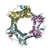
- PDB-2zvk: Crystal structure of PCNA in complex with DNA polymerase eta fragment -
+ Open data

- Basic information
| Entry | Database: PDB / ID: 2zvk | ||||||
|---|---|---|---|---|---|---|---|
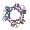
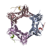
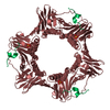
| Title | Crystal structure of PCNA in complex with DNA polymerase eta fragment | ||||||
 Components Components |
| ||||||
 Keywords Keywords | TRANSFERASE / DNA replication / PCNA / clamp / translesion synthesis / TLS / DNA polymerase / TLS polymerase / complex / PIP-box / DNA polymerase eta / DNA-binding / Nucleus / Systemic lupus erythematosus / Disease mutation / DNA damage / DNA repair / DNA synthesis / DNA-directed DNA polymerase / Magnesium / Metal-binding / Mutator protein / Nucleotidyltransferase / Phosphoprotein / Schiff base / Xeroderma pigmentosum | ||||||
| Function / homology |  Function and homology information Function and homology informationdinucleotide insertion or deletion binding / PCNA-p21 complex / mitotic telomere maintenance via semi-conservative replication / purine-specific mismatch base pair DNA N-glycosylase activity / nuclear lamina / Polymerase switching / Processive synthesis on the lagging strand / PCNA complex / MutLalpha complex binding / Telomere C-strand (Lagging Strand) Synthesis ...dinucleotide insertion or deletion binding / PCNA-p21 complex / mitotic telomere maintenance via semi-conservative replication / purine-specific mismatch base pair DNA N-glycosylase activity / nuclear lamina / Polymerase switching / Processive synthesis on the lagging strand / PCNA complex / MutLalpha complex binding / Telomere C-strand (Lagging Strand) Synthesis / Removal of the Flap Intermediate / Mismatch repair (MMR) directed by MSH2:MSH3 (MutSbeta) / Mismatch repair (MMR) directed by MSH2:MSH6 (MutSalpha) / Transcription of E2F targets under negative control by DREAM complex / Polymerase switching on the C-strand of the telomere / Processive synthesis on the C-strand of the telomere / replisome / Removal of the Flap Intermediate from the C-strand / response to L-glutamate / response to dexamethasone / histone acetyltransferase binding / G1/S-Specific Transcription / leading strand elongation / DNA polymerase processivity factor activity / nuclear replication fork / SUMOylation of DNA replication proteins / replication fork processing / PCNA-Dependent Long Patch Base Excision Repair / response to cadmium ion / estrous cycle / mismatch repair / cyclin-dependent protein kinase holoenzyme complex / base-excision repair, gap-filling / translesion synthesis / DNA polymerase binding / epithelial cell differentiation / TP53 Regulates Transcription of Genes Involved in G2 Cell Cycle Arrest / liver regeneration / positive regulation of DNA replication / nuclear estrogen receptor binding / replication fork / positive regulation of DNA repair / Translesion synthesis by REV1 / Translesion synthesis by POLK / male germ cell nucleus / Translesion synthesis by POLI / Gap-filling DNA repair synthesis and ligation in GG-NER / Termination of translesion DNA synthesis / Translesion Synthesis by POLH / Recognition of DNA damage by PCNA-containing replication complex / receptor tyrosine kinase binding / cellular response to xenobiotic stimulus / HDR through Homologous Recombination (HRR) / Dual Incision in GG-NER / cellular response to hydrogen peroxide / Dual incision in TC-NER / Gap-filling DNA repair synthesis and ligation in TC-NER / cellular response to UV / response to estradiol / E3 ubiquitin ligases ubiquitinate target proteins / heart development / chromatin organization / damaged DNA binding / chromosome, telomeric region / nuclear body / chromatin binding / centrosome / chromatin / protein-containing complex binding / enzyme binding / negative regulation of transcription by RNA polymerase II / extracellular exosome / nucleoplasm / identical protein binding / nucleus Similarity search - Function | ||||||
| Biological species |  Homo sapiens (human) Homo sapiens (human) | ||||||
| Method |  X-RAY DIFFRACTION / SYNCHROTRON / MOLECULAR REPLACEMENT / Resolution: 2.7 Å X-RAY DIFFRACTION / SYNCHROTRON / MOLECULAR REPLACEMENT / Resolution: 2.7 Å | ||||||
 Authors Authors | Hishiki, A. / Hashimoto, H. / Hanafusa, T. / Kamei, K. / Ohashi, E. / Shimizu, T. / Ohmori, H. / Sato, M. | ||||||
 Citation Citation | Journal: J.Biol.Chem. / Year: 2009 Title: Structural Basis for Novel Interactions between Human Translesion Synthesis Polymerases and Proliferating Cell Nuclear Antigen Authors: Hishiki, A. / Hashimoto, H. / Hanafusa, T. / Kamei, K. / Ohashi, E. / Shimizu, T. / Ohmori, H. / Sato, M. | ||||||
| History |
|
- Structure visualization
| Structure viewer | Molecule: MolmilJmol/JSmol |
|---|
- Downloads & links
-Download
| PDBx/mmCIF format | 2zvk.cif.gz | 158.1 KB | Display | PDBx/mmCIF format |
|---|---|---|---|---|
| PDB format | pdb2zvk.ent.gz | 125.9 KB | Display | PDB format |
| PDBx/mmJSON format | 2zvk.json.gz | Tree view | PDBx/mmJSON format | |
| Others |  Other downloads Other downloads |
-Validation report
| Arichive directory | https://data.pdbj.org/pub/pdb/validation_reports/zv/2zvkftp://data.pdbj.org/pub/pdb/validation_reports/zv/2zvk | HTTPS FTP |
|---|
-Related structure data
| Related structure data |  2zvlC  2zvmC 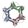 1vymS C: citing same article ( S: Starting model for refinement |
|---|---|
| Similar structure data |


-Links
 PDBj
PDBj


- Assembly
| Deposited unit | 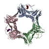
| ||||||||
|---|---|---|---|---|---|---|---|---|---|
| 1 |
| ||||||||
| Unit cell |
|
-Components
| #1: Protein | Mass: 28795.752 Da / Num. of mol.: 3 Source method: isolated from a genetically manipulated source Source: (gene. exp.) Homo sapiens (human) / Gene: PCNA / Plasmid: pT7 / Production host:  #2: Protein/peptide | Mass: 2507.949 Da / Num. of mol.: 3 / Source method: obtained synthetically / Details: chemically synthesized peptide / References: DNA-directed DNA polymerase #3: Water | ChemComp-HOH / |  Mass: 18.015 Da / Num. of mol.: 18 / Source method: isolated from a natural source / Formula: H2O Mass: 18.015 Da / Num. of mol.: 18 / Source method: isolated from a natural source / Formula: H2OHas protein modification | Y | |
|---|
-Experimental details
-Experiment
| Experiment | Method: X-RAY DIFFRACTION / Number of used crystals: 1 |
|---|
- Sample preparation
| Crystal | Density Matthews: 2.78 Å3/Da / Density % sol: 55.76 % |
|---|---|
| Crystal grow | Temperature: 293 K / pH: 3.6 Details: pH3.6, HANGING DROP VAPOR DIFFUSION, temperature 293K |
-Data collection
| Diffraction | Mean temperature: 100 K |
|---|---|
| Diffraction source | Source: SYNCHROTRON / Site: Photon Factory  / Beamline: AR-NW12A / Wavelength: 1 Å / Beamline: AR-NW12A / Wavelength: 1 Å |
| Detector | Type: ADSC QUANTUM 210 / Detector: CCD / Date: Oct 19, 2005 / Details: Rhodium coated silicon single crystal mirrors |
| Radiation | Monochromator: Numerical link type Si(111) double crystal monochromator, liquid nitrogen cooling Protocol: SINGLE WAVELENGTH / Monochromatic (M) / Laue (L): M / Scattering type: x-ray |
| Radiation wavelength | Wavelength: 1 Å / Relative weight: 1 |
| Reflection | Resolution: 2.7→50 Å / Num. obs: 27933 / % possible obs: 92.1 % / Observed criterion σ(I): -0.4 / Redundancy: 2.5 % / Biso Wilson estimate: 59 Å2 / Rmerge(I) obs: 0.093 / Net I/σ(I): 8.1 |
| Reflection shell | Resolution: 2.7→2.8 Å / Redundancy: 2 % / Rmerge(I) obs: 0.328 / Mean I/σ(I) obs: 2.6 / % possible all: 81.3 |
- Processing
| Software |
| |||||||||||||||||||||||||||||||||||||||||||||||||||||||||||||||||||||||||||||||||||||||||||||||||||||||||||||||||||||||||||||||||||||||||||||||||||||||||||||||||||||||||||||||
|---|---|---|---|---|---|---|---|---|---|---|---|---|---|---|---|---|---|---|---|---|---|---|---|---|---|---|---|---|---|---|---|---|---|---|---|---|---|---|---|---|---|---|---|---|---|---|---|---|---|---|---|---|---|---|---|---|---|---|---|---|---|---|---|---|---|---|---|---|---|---|---|---|---|---|---|---|---|---|---|---|---|---|---|---|---|---|---|---|---|---|---|---|---|---|---|---|---|---|---|---|---|---|---|---|---|---|---|---|---|---|---|---|---|---|---|---|---|---|---|---|---|---|---|---|---|---|---|---|---|---|---|---|---|---|---|---|---|---|---|---|---|---|---|---|---|---|---|---|---|---|---|---|---|---|---|---|---|---|---|---|---|---|---|---|---|---|---|---|---|---|---|---|---|---|---|---|
| Refinement | Method to determine structure: MOLECULAR REPLACEMENT Starting model: PDB ENTRY 1VYM Resolution: 2.7→20 Å / Cor.coef. Fo:Fc: 0.939 / Cor.coef. Fo:Fc free: 0.88 / SU B: 33.735 / SU ML: 0.3 / TLS residual ADP flag: LIKELY RESIDUAL / Cross valid method: THROUGHOUT / ESU R: 0.776 / ESU R Free: 0.367 / Stereochemistry target values: MAXIMUM LIKELIHOOD / Details: HYDROGENS HAVE BEEN ADDED IN THE RIDING POSITIONS
| |||||||||||||||||||||||||||||||||||||||||||||||||||||||||||||||||||||||||||||||||||||||||||||||||||||||||||||||||||||||||||||||||||||||||||||||||||||||||||||||||||||||||||||||
| Solvent computation | Ion probe radii: 0.8 Å / Shrinkage radii: 0.8 Å / VDW probe radii: 1.4 Å / Solvent model: BABINET MODEL WITH MASK | |||||||||||||||||||||||||||||||||||||||||||||||||||||||||||||||||||||||||||||||||||||||||||||||||||||||||||||||||||||||||||||||||||||||||||||||||||||||||||||||||||||||||||||||
| Displacement parameters | Biso mean: 30.089 Å2
| |||||||||||||||||||||||||||||||||||||||||||||||||||||||||||||||||||||||||||||||||||||||||||||||||||||||||||||||||||||||||||||||||||||||||||||||||||||||||||||||||||||||||||||||
| Refinement step | Cycle: LAST / Resolution: 2.7→20 Å
| |||||||||||||||||||||||||||||||||||||||||||||||||||||||||||||||||||||||||||||||||||||||||||||||||||||||||||||||||||||||||||||||||||||||||||||||||||||||||||||||||||||||||||||||
| Refine LS restraints |
| |||||||||||||||||||||||||||||||||||||||||||||||||||||||||||||||||||||||||||||||||||||||||||||||||||||||||||||||||||||||||||||||||||||||||||||||||||||||||||||||||||||||||||||||
| LS refinement shell | Resolution: 2.701→2.77 Å / Total num. of bins used: 20
| |||||||||||||||||||||||||||||||||||||||||||||||||||||||||||||||||||||||||||||||||||||||||||||||||||||||||||||||||||||||||||||||||||||||||||||||||||||||||||||||||||||||||||||||
| Refinement TLS params. | Method: refined / Refine-ID: X-RAY DIFFRACTION
| |||||||||||||||||||||||||||||||||||||||||||||||||||||||||||||||||||||||||||||||||||||||||||||||||||||||||||||||||||||||||||||||||||||||||||||||||||||||||||||||||||||||||||||||
| Refinement TLS group |
|
