[English] 日本語
Yorodumi
- PDB-1u76: Crystal structure of hPCNA bound to residues 452-466 of the DNA p... -

+
Open data


ID or keywords:

Loading...

-
Basic information

Entry
Database: PDB / ID: 1u76
Title
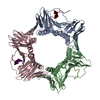
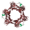
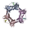
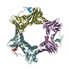
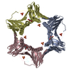
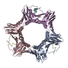
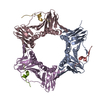
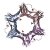
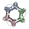
Crystal structure of hPCNA bound to residues 452-466 of the DNA polymerase-delta-p66 subunit
Components
  • KANRQVSITGFFQRK peptide from DNA polymerase delta subunit 3
  • Proliferating cell nuclear antigen
KeywordsREPLICATION / DNA replication / sliding clamp / DNA polymerase delta / p66 / PIP-Box
Function / homology
Function and homology information


delta DNA polymerase complex / DNA synthesis involved in UV-damage excision repair / zeta DNA polymerase complex / positive regulation of deoxyribonuclease activity / dinucleotide insertion or deletion binding / PCNA-p21 complex / mitotic telomere maintenance via semi-conservative replication / purine-specific mismatch base pair DNA N-glycosylase activity / nuclear lamina / MutLalpha complex binding ...delta DNA polymerase complex / DNA synthesis involved in UV-damage excision repair / zeta DNA polymerase complex / positive regulation of deoxyribonuclease activity / dinucleotide insertion or deletion binding / PCNA-p21 complex / mitotic telomere maintenance via semi-conservative replication / purine-specific mismatch base pair DNA N-glycosylase activity / nuclear lamina / MutLalpha complex binding / positive regulation of DNA-directed DNA polymerase activity / Polymerase switching / Telomere C-strand (Lagging Strand) Synthesis / Processive synthesis on the lagging strand / PCNA complex / Removal of the Flap Intermediate / Processive synthesis on the C-strand of the telomere / Mismatch repair (MMR) directed by MSH2:MSH3 (MutSbeta) / Polymerase switching on the C-strand of the telomere / Mismatch repair (MMR) directed by MSH2:MSH6 (MutSalpha) / Transcription of E2F targets under negative control by DREAM complex / Removal of the Flap Intermediate from the C-strand / replisome / nucleotide-excision repair, DNA gap filling / response to L-glutamate / DNA strand elongation involved in DNA replication / histone acetyltransferase binding / DNA synthesis involved in DNA repair / DNA polymerase processivity factor activity / DNA biosynthetic process / G1/S-Specific Transcription / leading strand elongation / response to dexamethasone / replication fork processing / nuclear replication fork / SUMOylation of DNA replication proteins / estrous cycle / PCNA-Dependent Long Patch Base Excision Repair / cyclin-dependent protein kinase holoenzyme complex / error-prone translesion synthesis / mismatch repair / translesion synthesis / response to cadmium ion / DNA polymerase binding / epithelial cell differentiation / positive regulation of DNA repair / Translesion synthesis by REV1 / Translesion synthesis by POLK / Translesion synthesis by POLI / Gap-filling DNA repair synthesis and ligation in GG-NER / base-excision repair, gap-filling / TP53 Regulates Transcription of Genes Involved in G2 Cell Cycle Arrest / positive regulation of DNA replication / replication fork / male germ cell nucleus / liver regeneration / nuclear estrogen receptor binding / Recognition of DNA damage by PCNA-containing replication complex / Termination of translesion DNA synthesis / Translesion Synthesis by POLH / HDR through Homologous Recombination (HRR) / Dual Incision in GG-NER / DNA-templated DNA replication / receptor tyrosine kinase binding / cellular response to hydrogen peroxide / Dual incision in TC-NER / Gap-filling DNA repair synthesis and ligation in TC-NER / cellular response to UV / cellular response to xenobiotic stimulus / E3 ubiquitin ligases ubiquitinate target proteins / response to estradiol / protein-macromolecule adaptor activity / heart development / chromosome, telomeric region / damaged DNA binding / DNA-directed DNA polymerase activity / nuclear body / centrosome / chromatin binding / protein-containing complex binding / chromatin / enzyme binding / negative regulation of transcription by RNA polymerase II / extracellular exosome / nucleoplasm / identical protein binding / nucleus / cytoplasm
Similarity search - Function
DNA polymerase delta subunit 3 / DNA polymerase delta subunit 3 superfamily / DNA polymerase subunit Cdc27 / Box / Proliferating Cell Nuclear Antigen / Proliferating Cell Nuclear Antigen - #10 / Proliferating cell nuclear antigen signature 2. / Proliferating cell nuclear antigen, PCNA, conserved site / Proliferating cell nuclear antigen signature 1. / Proliferating cell nuclear antigen, PCNA ...DNA polymerase delta subunit 3 / DNA polymerase delta subunit 3 superfamily / DNA polymerase subunit Cdc27 / Box / Proliferating Cell Nuclear Antigen / Proliferating Cell Nuclear Antigen - #10 / Proliferating cell nuclear antigen signature 2. / Proliferating cell nuclear antigen, PCNA, conserved site / Proliferating cell nuclear antigen signature 1. / Proliferating cell nuclear antigen, PCNA / Proliferating cell nuclear antigen, PCNA, N-terminal / Proliferating cell nuclear antigen, PCNA, C-terminal / Proliferating cell nuclear antigen, N-terminal domain / Proliferating cell nuclear antigen, C-terminal domain / : / Alpha Beta
Similarity search - Domain/homology
Proliferating cell nuclear antigen / DNA polymerase delta subunit 3
Similarity search - Component
Biological speciesHomo sapiens (human)
MethodX-RAY DIFFRACTION / SYNCHROTRON / MOLECULAR REPLACEMENT / Resolution: 2.6 Å
AuthorsBruning, J.B. / Shamoo, Y.
CitationJournal: Structure / Year: 2004
Title: Structural and Thermodynamic Analysis of Human PCNA with Peptides Derived from DNA Polymerase-delta p66 Subunit and Flap Endonuclease-1.
Authors: Bruning, J.B. / Shamoo, Y.
History
DepositionAug 2, 2004Deposition site: RCSB / Processing site: RCSB
Revision 1.0Dec 14, 2004Provider: repository / Type: Initial release
Revision 1.1Apr 30, 2008Group: Version format compliance
Revision 1.2Jul 13, 2011Group: Version format compliance
Revision 1.3Aug 23, 2023Group: Data collection / Database references / Refinement description
Category: chem_comp_atom / chem_comp_bond ...chem_comp_atom / chem_comp_bond / database_2 / pdbx_initial_refinement_model
Item: _database_2.pdbx_DOI / _database_2.pdbx_database_accession

-
Structure visualization

Structure viewerMolecule:
MolmilJmol/JSmol

Downloads & links

-
Assembly

Deposited unit
A: Proliferating cell nuclear antigen
B: KANRQVSITGFFQRK peptide from DNA polymerase delta subunit 3
C: Proliferating cell nuclear antigen
D: KANRQVSITGFFQRK peptide from DNA polymerase delta subunit 3
E: Proliferating cell nuclear antigen
F: KANRQVSITGFFQRK peptide from DNA polymerase delta subunit 3


Theoretical massNumber of molelcules
Total (without water)91,7396
Polymers91,7396
Non-polymers00
Water10,827601
1


  • Idetical with deposited unit
  • defined by author&software
TypeNameSymmetry operationNumber
identity operation1_555x,y,z1
Buried area8920 Å2
ΔGint-53 kcal/mol
Surface area33750 Å2
MethodPISA
Unit cell
Length a, b, c (Å)82.480, 82.480, 203.800
Angle α, β, γ (deg.)90.00, 90.00, 120.00
Int Tables number154
Space group name H-MP3221

-
Components

#1: Protein Proliferating cell nuclear antigen / PCNA / Cyclin


Mass: 28795.752 Da / Num. of mol.: 3
Source method: isolated from a genetically manipulated source
Source: (gene. exp.) Homo sapiens (human) / Gene: PCNA / Plasmid: pET28a-hPCNA / Species (production host): Escherichia coli / Production host: Escherichia coli BL21(DE3) (bacteria) / Strain (production host): BL21DE3 / References: UniProt: P12004
#2: Protein/peptide KANRQVSITGFFQRK peptide from DNA polymerase delta subunit 3


Mass: 1784.070 Da / Num. of mol.: 3 / Fragment: PIP-box region of p66 (residues 452-466) / Source method: obtained synthetically
Details: the peptide was chemically synthesized, the sequence of the peptide is naturally found in Homo sapiens (Human)
References: UniProt: Q15054
#3: Water ChemComp-HOH / water


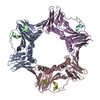
Mass: 18.015 Da / Num. of mol.: 601 / Source method: isolated from a natural source / Formula: H2O

-
Experimental details

-
Experiment

ExperimentMethod: X-RAY DIFFRACTION / Number of used crystals: 1

-
Sample preparation

CrystalDensity Matthews: 2.328 Å3/Da / Density % sol: 59.01 %
Crystal growTemperature: 298 K / Method: vapor diffusion, hanging drop / pH: 4.6
Details: ammonium sulfate, sodium acetate, pH 4.6, VAPOR DIFFUSION, HANGING DROP, temperature 298K

-
Data collection

DiffractionMean temperature: 100 K
Diffraction sourceSource: SYNCHROTRON / Site: CHESS / Beamline: A1 / Wavelength: 0.9764 Å
DetectorType: ADSC QUANTUM 4 / Detector: CCD / Date: Feb 29, 2004
RadiationMonochromator: Si 111 CHANNEL / Protocol: SINGLE WAVELENGTH / Monochromatic (M) / Laue (L): M / Scattering type: x-ray
Radiation wavelengthWavelength: 0.9764 Å / Relative weight: 1
ReflectionResolution: 2.6→20 Å / Num. all: 24477 / Num. obs: 24477 / % possible obs: 96 % / Observed criterion σ(F): 0 / Observed criterion σ(I): 0 / Redundancy: 4.36 % / Biso Wilson estimate: 57.4 Å2 / Rmerge(I) obs: 0.082 / Rsym value: 0.082 / Net I/σ(I): 7.4
Reflection shellResolution: 2.6→2.69 Å / Redundancy: 5.1 % / Rmerge(I) obs: 0.336 / Mean I/σ(I) obs: 3.8 / Num. unique all: 2482 / Rsym value: 0.336 / % possible all: 100

-
Processing

Software
NameVersionClassification
CNS1.1refinement
DENZOdata reduction
SCALEPACKdata scaling
CNSphasing
RefinementMethod to determine structure: MOLECULAR REPLACEMENT
Starting model: PDB 1AXC.pdb without chains B,D,F
Resolution: 2.6→10 Å / Rfactor Rfree error: 0.008 / Data cutoff high absF: 428998.24 / Data cutoff low absF: 0 / Isotropic thermal model: RESTRAINED / Cross valid method: THROUGHOUT / σ(F): 2 / σ(I): 0 / Stereochemistry target values: Engh & Huber
RfactorNum. reflection% reflectionSelection details
Rfree0.278 1150 4.9 %RANDOM
Rwork0.24 ---
obs0.24 23519 94 %-
all-23519 --
Solvent computationSolvent model: FLAT MODEL / Bsol: 72.8016 Å2 / ksol: 0.401949 e/Å3
Displacement parametersBiso mean: 65.2 Å2
Baniso -1Baniso -2Baniso -3
1--12.5 Å26.43 Å20 Å2
2---12.5 Å20 Å2
3---25.01 Å2
Refine analyze
FreeObs
Luzzati coordinate error0.44 Å0.37 Å
Luzzati d res low-10 Å
Luzzati sigma a0.57 Å0.49 Å
Refinement stepCycle: LAST / Resolution: 2.6→10 Å
ProteinNucleic acidLigandSolventTotal
Num. atoms6100 0 0 601 6701
Refine LS restraints
Refine-IDTypeDev idealDev ideal target
X-RAY DIFFRACTIONc_bond_d0.007
X-RAY DIFFRACTIONc_bond_d_na
X-RAY DIFFRACTIONc_bond_d_prot
X-RAY DIFFRACTIONc_angle_d
X-RAY DIFFRACTIONc_angle_d_na
X-RAY DIFFRACTIONc_angle_d_prot
X-RAY DIFFRACTIONc_angle_deg1.3
X-RAY DIFFRACTIONc_angle_deg_na
X-RAY DIFFRACTIONc_angle_deg_prot
X-RAY DIFFRACTIONc_dihedral_angle_d25.4
X-RAY DIFFRACTIONc_dihedral_angle_d_na
X-RAY DIFFRACTIONc_dihedral_angle_d_prot
X-RAY DIFFRACTIONc_improper_angle_d0.73
X-RAY DIFFRACTIONc_improper_angle_d_na
X-RAY DIFFRACTIONc_improper_angle_d_prot
X-RAY DIFFRACTIONc_mcbond_it3.033
X-RAY DIFFRACTIONc_mcangle_it4.844
X-RAY DIFFRACTIONc_scbond_it7.226
X-RAY DIFFRACTIONc_scangle_it9.417.5
LS refinement shellResolution: 2.6→2.69 Å / Rfactor Rfree error: 0.038 / Total num. of bins used: 10
RfactorNum. reflection% reflection
Rfree0.423 124 5.2 %
Rwork0.418 2248 -
obs-2372 97.4 %
Xplor file
Refine-IDSerial noParam fileTopol file
X-RAY DIFFRACTION1PROTEIN_REP.PARAMPROTEIN.TOP
X-RAY DIFFRACTION2WATER_REP.PARAMWATER.TOP

+
About Yorodumi

-
News

-
Feb 9, 2022. New format data for meta-information of EMDB entries

New format data for meta-information of EMDB entries

  • Version 3 of the EMDB header file is now the official format.
  • The previous official version 1.9 will be removed from the archive.

Related info.:EMDB header

External links:wwPDB to switch to version 3 of the EMDB data model

-
Aug 12, 2020. Covid-19 info

Covid-19 info

URL: https://pdbjlvh1.pdbj.org/emnavi/covid19.php

New page: Covid-19 featured information page in EM Navigator.

Related info.:Covid-19 info / Mar 5, 2020. Novel coronavirus structure data

+
Mar 5, 2020. Novel coronavirus structure data

Novel coronavirus structure data

Related info.:Yorodumi Speices / Aug 12, 2020. Covid-19 info

External links:COVID-19 featured content - PDBj / Molecule of the Month (242):Coronavirus Proteases

+
Jan 31, 2019. EMDB accession codes are about to change! (news from PDBe EMDB page)

EMDB accession codes are about to change! (news from PDBe EMDB page)

  • The allocation of 4 digits for EMDB accession codes will soon come to an end. Whilst these codes will remain in use, new EMDB accession codes will include an additional digit and will expand incrementally as the available range of codes is exhausted. The current 4-digit format prefixed with “EMD-” (i.e. EMD-XXXX) will advance to a 5-digit format (i.e. EMD-XXXXX), and so on. It is currently estimated that the 4-digit codes will be depleted around Spring 2019, at which point the 5-digit format will come into force.
  • The EM Navigator/Yorodumi systems omit the EMD- prefix.

Related info.:Q: What is EMD? / ID/Accession-code notation in Yorodumi/EM Navigator

External links:EMDB Accession Codes are Changing Soon! / Contact to PDBj

+
Jul 12, 2017. Major update of PDB

Major update of PDB

  • wwPDB released updated PDB data conforming to the new PDBx/mmCIF dictionary.
  • This is a major update changing the version number from 4 to 5, and with Remediation, in which all the entries are updated.
  • In this update, many items about electron microscopy experimental information are reorganized (e.g. em_software).
  • Now, EM Navigator and Yorodumi are based on the updated data.

External links:wwPDB Remediation / Enriched Model Files Conforming to OneDep Data Standards Now Available in the PDB FTP Archive

-
Yorodumi

Thousand views of thousand structures

  • Yorodumi is a browser for structure data from EMDB, PDB, SASBDB, etc.
  • This page is also the successor to EM Navigator detail page, and also detail information page/front-end page for Omokage search.
  • The word "yorodu" (or yorozu) is an old Japanese word meaning "ten thousand". "mi" (miru) is to see.

Related info.:EMDB / PDB / SASBDB / Comparison of 3 databanks / Yorodumi Search / Aug 31, 2016. New EM Navigator & Yorodumi / Yorodumi Papers / Jmol/JSmol / Function and homology information / Changes in new EM Navigator and Yorodumi

Read more