 Movie
Movie Controller


+ Open data

- Basic information
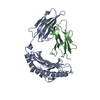
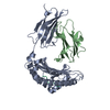
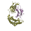
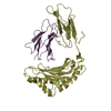
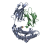
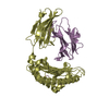
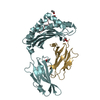
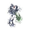
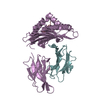
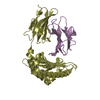
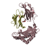
| Entry | Database: PDB / ID: 2zsv | ||||||
|---|---|---|---|---|---|---|---|
| Title | Crystal structure of H-2Kb in complex with JHMV epitope S598 | ||||||
 Components Components |
| ||||||
 Keywords Keywords | IMMUNE SYSTEM / Ig Fold / protein-protein interactions / subdominant epitope / Glycoprotein / Immune response / Membrane / MHC I / Transmembrane / Immunoglobulin domain / Secreted | ||||||
| Function / homology |  Function and homology information Function and homology informationMHC class Ib protein complex / natural killer cell lectin-like receptor binding / TAP2 binding / TAP1 binding / cis-Golgi network membrane / Endosomal/Vacuolar pathway / DAP12 interactions / Antigen Presentation: Folding, assembly and peptide loading of class I MHC / ER-Phagosome pathway / DAP12 signaling ...MHC class Ib protein complex / natural killer cell lectin-like receptor binding / TAP2 binding / TAP1 binding / cis-Golgi network membrane / Endosomal/Vacuolar pathway / DAP12 interactions / Antigen Presentation: Folding, assembly and peptide loading of class I MHC / ER-Phagosome pathway / DAP12 signaling / Immunoregulatory interactions between a Lymphoid and a non-Lymphoid cell / antigen processing and presentation of endogenous peptide antigen via MHC class I via ER pathway, TAP-dependent / TAP complex binding / antigen processing and presentation of exogenous peptide antigen via MHC class I / Golgi medial cisterna / regulation of membrane depolarization / inner ear development / CD8 receptor binding / endoplasmic reticulum exit site / TAP binding / beta-2-microglobulin binding / MHC class I protein binding / antigen processing and presentation of endogenous peptide antigen via MHC class Ib / antigen processing and presentation of endogenous peptide antigen via MHC class I via ER pathway, TAP-independent / cellular defense response / T cell receptor binding / Neutrophil degranulation / 14-3-3 protein binding / lumenal side of endoplasmic reticulum membrane / regulation of iron ion transport / cellular response to iron(III) ion / antigen processing and presentation of exogenous protein antigen via MHC class Ib, TAP-dependent / negative regulation of iron ion transport / negative regulation of forebrain neuron differentiation / regulation of erythrocyte differentiation / iron ion transport / peptide antigen assembly with MHC class I protein complex / response to molecule of bacterial origin / HFE-transferrin receptor complex / MHC class I peptide loading complex / transferrin transport / negative regulation of receptor-mediated endocytosis / cellular response to iron ion / positive regulation of T cell cytokine production / antigen processing and presentation of endogenous peptide antigen via MHC class I / MHC class I protein complex / peptide antigen assembly with MHC class II protein complex / negative regulation of neurogenesis / multicellular organismal-level iron ion homeostasis / cellular response to nicotine / MHC class II protein complex / positive regulation of receptor-mediated endocytosis / positive regulation of T cell mediated cytotoxicity / negative regulation of epithelial cell proliferation / antigen processing and presentation of exogenous peptide antigen via MHC class II / positive regulation of immune response / peptide antigen binding / phagocytic vesicle membrane / positive regulation of T cell activation / sensory perception of smell / positive regulation of cellular senescence / MHC class II protein complex binding / T cell differentiation in thymus / late endosome membrane / negative regulation of neuron projection development / antimicrobial humoral immune response mediated by antimicrobial peptide / antibacterial humoral response / cellular response to lipopolysaccharide / protein refolding / protein-folding chaperone binding / early endosome membrane / amyloid fibril formation / defense response to Gram-negative bacterium / protein homotetramerization / intracellular iron ion homeostasis / host cell endoplasmic reticulum-Golgi intermediate compartment membrane / learning or memory / early endosome / receptor-mediated virion attachment to host cell / defense response to bacterium / defense response to Gram-positive bacterium / immune response / endocytosis involved in viral entry into host cell / receptor ligand activity / Golgi membrane / fusion of virus membrane with host plasma membrane / external side of plasma membrane / signaling receptor binding / innate immune response / lysosomal membrane / fusion of virus membrane with host endosome membrane / viral envelope / host cell plasma membrane / virion membrane / structural molecule activity / Golgi apparatus / cell surface / endoplasmic reticulum / protein homodimerization activity / : Similarity search - Function | ||||||
| Biological species |  | ||||||
| Method |  X-RAY DIFFRACTION / MOLECULAR REPLACEMENT / Resolution: 1.8 Å X-RAY DIFFRACTION / MOLECULAR REPLACEMENT / Resolution: 1.8 Å | ||||||
 Authors Authors | Theodossis, A. / Dunstone, M.A. / Rossjohn, J. | ||||||
 Citation Citation | Journal: Plos Pathog. / Year: 2008 Title: Prevention of cytotoxic T cell escape using a heteroclitic subdominant viral T cell determinant. Authors: Butler, N.S. / Theodossis, A. / Webb, A.I. / Nastovska, R. / Ramarathinam, S.H. / Dunstone, M.A. / Rossjohn, J. / Purcell, A.W. / Perlman, S. | ||||||
| History |
|
- Structure visualization
| Structure viewer | Molecule: MolmilJmol/JSmol |
|---|
- Downloads & links
-Download
| PDBx/mmCIF format | 2zsv.cif.gz | 183 KB | Display | PDBx/mmCIF format |
|---|---|---|---|---|
| PDB format | pdb2zsv.ent.gz | 144.6 KB | Display | PDB format |
| PDBx/mmJSON format | 2zsv.json.gz | Tree view | PDBx/mmJSON format | |
| Others |  Other downloads Other downloads |
-Validation report
| Arichive directory | https://data.pdbj.org/pub/pdb/validation_reports/zs/2zsvftp://data.pdbj.org/pub/pdb/validation_reports/zs/2zsv | HTTPS FTP |
|---|
-Related structure data
| Related structure data | 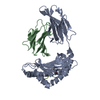 2zswC 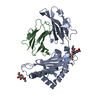 1g7qS C: citing same article ( S: Starting model for refinement |
|---|---|
| Similar structure data |


-Links
 PDBj
PDBj


- Assembly
| Deposited unit | 
| |||||||||||||||||||||||||||||||||||||||||||||||||||||||||||||||||||||||||||||||||||||||||||||||||||
|---|---|---|---|---|---|---|---|---|---|---|---|---|---|---|---|---|---|---|---|---|---|---|---|---|---|---|---|---|---|---|---|---|---|---|---|---|---|---|---|---|---|---|---|---|---|---|---|---|---|---|---|---|---|---|---|---|---|---|---|---|---|---|---|---|---|---|---|---|---|---|---|---|---|---|---|---|---|---|---|---|---|---|---|---|---|---|---|---|---|---|---|---|---|---|---|---|---|---|---|---|
| 1 | 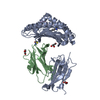
| |||||||||||||||||||||||||||||||||||||||||||||||||||||||||||||||||||||||||||||||||||||||||||||||||||
| 2 | 
| |||||||||||||||||||||||||||||||||||||||||||||||||||||||||||||||||||||||||||||||||||||||||||||||||||
| Unit cell |
| |||||||||||||||||||||||||||||||||||||||||||||||||||||||||||||||||||||||||||||||||||||||||||||||||||
| Noncrystallographic symmetry (NCS) | NCS domain:
NCS domain segments: Component-ID: 1 / Refine code: 5
NCS ensembles :
|
-Components
-Protein , 2 types, 4 molecules ACBD
| #1: Protein | Mass: 32068.777 Da / Num. of mol.: 2 / Fragment: extracellular domain Source method: isolated from a genetically manipulated source Source: (gene. exp.)  #2: Protein | Mass: 11704.359 Da / Num. of mol.: 2 Source method: isolated from a genetically manipulated source Source: (gene. exp.) |
|---|
-Protein/peptide , 1 types, 2 molecules EF
| #3: Protein/peptide | Mass: 947.113 Da / Num. of mol.: 2 / Source method: obtained synthetically Details: The peptide was chemically synthesized. The sequence of the peptide is naturally found in Mouse Hepatitis Virus JHM strain References: UniProt: P11225*PLUS |
|---|
-Non-polymers , 3 types, 627 molecules 


| #4: Chemical | ChemComp-GOL /  Mass: 92.094 Da / Num. of mol.: 10 / Source method: obtained synthetically / Formula: C3H8O3 Mass: 92.094 Da / Num. of mol.: 10 / Source method: obtained synthetically / Formula: C3H8O3#5: Chemical | ChemComp-CYS / |  Type: L-peptide linking / Mass: 121.158 Da / Num. of mol.: 1 / Source method: obtained synthetically / Formula: C3H7NO2S Type: L-peptide linking / Mass: 121.158 Da / Num. of mol.: 1 / Source method: obtained synthetically / Formula: C3H7NO2S#6: Water | ChemComp-HOH / | Mass: 18.015 Da / Num. of mol.: 616 / Source method: isolated from a natural source / Formula: H2O |
|---|
-Experimental details
-Experiment
| Experiment | Method: X-RAY DIFFRACTION / Number of used crystals: 1 |
|---|
- Sample preparation
| Crystal | Density Matthews: 2.78 Å3/Da / Density % sol: 55.77 % |
|---|---|
| Crystal grow | Temperature: 294 K / Method: vapor diffusion, hanging drop / pH: 6.6 Details: 0.1M cacodylate, 16% PEG 8K, 0.2M Ca(OAc)2, pH 6.6, VAPOR DIFFUSION, HANGING DROP, temperature 294K |
-Data collection
| Diffraction | Mean temperature: 100 K |
|---|---|
| Diffraction source | Source: ROTATING ANODE / Type: RIGAKU / Wavelength: 1.54178 Å |
| Detector | Type: RIGAKU RAXIS IV++ / Detector: IMAGE PLATE / Date: Nov 8, 2005 |
| Radiation | Protocol: SINGLE WAVELENGTH / Monochromatic (M) / Laue (L): M / Scattering type: x-ray |
| Radiation wavelength | Wavelength: 1.54178 Å / Relative weight: 1 |
| Reflection | Resolution: 1.8→24.46 Å / Num. all: 90277 / Num. obs: 90277 / % possible obs: 99.6 % / Redundancy: 2.9 % / Biso Wilson estimate: 24.9 Å2 / Rmerge(I) obs: 0.046 / Net I/σ(I): 18 |
| Reflection shell | Resolution: 1.8→1.9 Å / Redundancy: 2.7 % / Rmerge(I) obs: 0.385 / Mean I/σ(I) obs: 2 / Num. unique all: 13114 / % possible all: 99.5 |
- Processing
| Software |
| |||||||||||||||||||||||||||||||||||||||||||||||||||||||||||||||||||||||||||||||||||||||||||
|---|---|---|---|---|---|---|---|---|---|---|---|---|---|---|---|---|---|---|---|---|---|---|---|---|---|---|---|---|---|---|---|---|---|---|---|---|---|---|---|---|---|---|---|---|---|---|---|---|---|---|---|---|---|---|---|---|---|---|---|---|---|---|---|---|---|---|---|---|---|---|---|---|---|---|---|---|---|---|---|---|---|---|---|---|---|---|---|---|---|---|---|---|
| Refinement | Method to determine structure: MOLECULAR REPLACEMENT Starting model: 1G7Q Resolution: 1.8→24.4 Å / Cor.coef. Fo:Fc: 0.957 / Cor.coef. Fo:Fc free: 0.935 / SU B: 2.785 / SU ML: 0.087 / Cross valid method: THROUGHOUT / ESU R: 0.133 / ESU R Free: 0.13 / Stereochemistry target values: MAXIMUM LIKELIHOOD
| |||||||||||||||||||||||||||||||||||||||||||||||||||||||||||||||||||||||||||||||||||||||||||
| Solvent computation | Ion probe radii: 0.8 Å / Shrinkage radii: 0.8 Å / VDW probe radii: 1.2 Å / Solvent model: MASK | |||||||||||||||||||||||||||||||||||||||||||||||||||||||||||||||||||||||||||||||||||||||||||
| Displacement parameters | Biso mean: 23.616 Å2
| |||||||||||||||||||||||||||||||||||||||||||||||||||||||||||||||||||||||||||||||||||||||||||
| Refinement step | Cycle: LAST / Resolution: 1.8→24.4 Å
| |||||||||||||||||||||||||||||||||||||||||||||||||||||||||||||||||||||||||||||||||||||||||||
| Refine LS restraints |
| |||||||||||||||||||||||||||||||||||||||||||||||||||||||||||||||||||||||||||||||||||||||||||
| Refine LS restraints NCS | Refine-ID: X-RAY DIFFRACTION
| |||||||||||||||||||||||||||||||||||||||||||||||||||||||||||||||||||||||||||||||||||||||||||
| LS refinement shell | Resolution: 1.8→1.846 Å / Total num. of bins used: 20
|
