 Movie
Movie Controller

[English] 日本語
 Yorodumi
- PDB-2zqq: Crystal structure of human AUH (3-methylglutaconyl-coa hydratase)... -
+ Open data

- Basic information
| Entry | Database: PDB / ID: 2zqq | ||||||
|---|---|---|---|---|---|---|---|
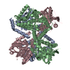
| Title | Crystal structure of human AUH (3-methylglutaconyl-coa hydratase) mixed with (AUUU)24A RNA | ||||||
 Components Components | Methylglutaconyl-CoA hydratase | ||||||
 Keywords Keywords | LYASE / BETA SPIRAL / Branched-chain amino acid catabolism / Disease mutation / Mitochondrion / RNA-binding / Transit peptide / Structural Genomics / NPPSFA / National Project on Protein Structural and Functional Analyses / RIKEN Structural Genomics/Proteomics Initiative / RSGI | ||||||
| Function / homology |  Function and homology information Function and homology informationmethylglutaconyl-CoA hydratase / itaconyl-CoA hydratase / itaconyl-CoA hydratase activity / 3-methylglutaconic aciduria / methylglutaconyl-CoA hydratase activity / L-leucine catabolic process / enoyl-CoA hydratase activity / Branched-chain amino acid catabolism / fatty acid beta-oxidation / mRNA 3'-UTR binding ...methylglutaconyl-CoA hydratase / itaconyl-CoA hydratase / itaconyl-CoA hydratase activity / 3-methylglutaconic aciduria / methylglutaconyl-CoA hydratase activity / L-leucine catabolic process / enoyl-CoA hydratase activity / Branched-chain amino acid catabolism / fatty acid beta-oxidation / mRNA 3'-UTR binding / mitochondrial matrix / mitochondrion Similarity search - Function | ||||||
| Biological species |  Homo sapiens (human) Homo sapiens (human) | ||||||
| Method |  X-RAY DIFFRACTION / SYNCHROTRON / MOLECULAR REPLACEMENT / Resolution: 2.2 Å X-RAY DIFFRACTION / SYNCHROTRON / MOLECULAR REPLACEMENT / Resolution: 2.2 Å | ||||||
 Authors Authors | Kurimoto, K. / Kuwasako, K. / Muto, Y. / Nureki, O. / Yokoyama, S. / RIKEN Structural Genomics/Proteomics Initiative (RSGI) | ||||||
 Citation Citation | Journal: Proteins / Year: 2009 Title: AU-rich RNA-binding induces changes in the quaternary structure of AUH Authors: Kurimoto, K. / Kuwasako, K. / Sandercock, A.M. / Unzai, S. / Robinson, C.V. / Muto, Y. / Yokoyama, S. | ||||||
| History |
|
- Structure visualization
| Structure viewer | Molecule: MolmilJmol/JSmol |
|---|
- Downloads & links
-Download
| PDBx/mmCIF format | 2zqq.cif.gz | 304.8 KB | Display | PDBx/mmCIF format |
|---|---|---|---|---|
| PDB format | pdb2zqq.ent.gz | 249.6 KB | Display | PDB format |
| PDBx/mmJSON format | 2zqq.json.gz | Tree view | PDBx/mmJSON format | |
| Others |  Other downloads Other downloads |
-Validation report
| Arichive directory | https://data.pdbj.org/pub/pdb/validation_reports/zq/2zqqftp://data.pdbj.org/pub/pdb/validation_reports/zq/2zqq | HTTPS FTP |
|---|
-Related structure data
| Related structure data |  2zqrC  1hzdS  1v4m 2e8k S: Starting model for refinement C: citing same article ( |
|---|---|
| Similar structure data | |
| Other databases |


-Links
 PDBj
PDBj- Assembly
| Deposited unit | 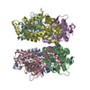
| ||||||||
|---|---|---|---|---|---|---|---|---|---|
| 1 |
| ||||||||
| Unit cell |
|
-Components
| #1: Protein | Mass: 29238.154 Da / Num. of mol.: 6 Source method: isolated from a genetically manipulated source Source: (gene. exp.) Homo sapiens (human) / Gene: AUH / Plasmid: PGEX6P1 / Production host:  #2: Water | ChemComp-HOH / |  Mass: 18.015 Da / Num. of mol.: 508 / Source method: isolated from a natural source / Formula: H2O Mass: 18.015 Da / Num. of mol.: 508 / Source method: isolated from a natural source / Formula: H2O |
|---|
-Experimental details
-Experiment
| Experiment | Method: X-RAY DIFFRACTION / Number of used crystals: 1 |
|---|
- Sample preparation
| Crystal | Density Matthews: 2.72 Å3/Da / Density % sol: 54.85 % |
|---|---|
| Crystal grow | Temperature: 298 K / Method: vapor diffusion, hanging drop / pH: 7.5 Details: DROP: 50MM HEPES, 1.0M AMMONIUM SULFATE, 1.0% PEG400, RESERVOIR: 100MM HEPES, 2.0M AMMONIUM SULFATE, 2.0% PEG400, pH 7.50, VAPOR DIFFUSION, HANGING DROP, temperature 298K |
-Data collection
| Diffraction | Mean temperature: 100 K |
|---|---|
| Diffraction source | Source: SYNCHROTRON / Site: SPring-8  / Beamline: BL41XU / Wavelength: 1 / Beamline: BL41XU / Wavelength: 1 |
| Detector | Detector: CCD / Date: May 23, 2002 |
| Radiation | Monochromator: SI111 / Protocol: SINGLE WAVELENGTH / Monochromatic (M) / Laue (L): M / Scattering type: x-ray |
| Radiation wavelength | Wavelength: 1 Å / Relative weight: 1 |
| Reflection | Resolution: 2.2→50 Å / Num. obs: 88656 / % possible obs: 92.8 % / Observed criterion σ(I): 2 / Redundancy: 2.29 % / Rmerge(I) obs: 0.071 / Rsym value: 0.071 / Net I/σ(I): 23.6 |
| Reflection shell | Resolution: 2.2→2.24 Å / Redundancy: 1.83 % / Rmerge(I) obs: 0.164 / Mean I/σ(I) obs: 4.725 / Rsym value: 0.156 / % possible all: 85.5 |
- Processing
| Software |
| ||||||||||||||||||||||||||||||||||||||||||||||||||||||||||||
|---|---|---|---|---|---|---|---|---|---|---|---|---|---|---|---|---|---|---|---|---|---|---|---|---|---|---|---|---|---|---|---|---|---|---|---|---|---|---|---|---|---|---|---|---|---|---|---|---|---|---|---|---|---|---|---|---|---|---|---|---|---|
| Refinement | Method to determine structure: MOLECULAR REPLACEMENT Starting model: 1HZD Resolution: 2.2→50 Å / σ(F): 0 / Stereochemistry target values: ENGH & HUBER
| ||||||||||||||||||||||||||||||||||||||||||||||||||||||||||||
| Refine analyze |
| ||||||||||||||||||||||||||||||||||||||||||||||||||||||||||||
| Refinement step | Cycle: LAST / Resolution: 2.2→50 Å
| ||||||||||||||||||||||||||||||||||||||||||||||||||||||||||||
| Refine LS restraints |
| ||||||||||||||||||||||||||||||||||||||||||||||||||||||||||||
| LS refinement shell | Resolution: 2.2→2.28 Å /
|
