 ムービー
ムービー コントローラー


+ データを開く

- 基本情報
基本情報
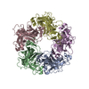
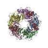
| 登録情報 | データベース: PDB / ID: 1hnu | ||||||
|---|---|---|---|---|---|---|---|
| タイトル | CRYSTAL STRUCTURE OF PEROXISOMAL DELTA3-DELTA2-ENOYL-COA ISOMERASE FROM SACCHAROMYCES CEREVISIAE | ||||||
 要素 要素 | D3,D2-ENOYL COA ISOMERASE ECI1 | ||||||
 キーワード キーワード | ISOMERASE / alpha/beta | ||||||
| 機能・相同性 |  機能・相同性情報 機能・相同性情報Beta-oxidation of very long chain fatty acids / Delta3-Delta2-enoyl-CoA isomerase / delta(3)-delta(2)-enoyl-CoA isomerase activity / Peroxisomal protein import / fatty acid beta-oxidation / peroxisomal matrix / peroxisome 類似検索 - 分子機能 | ||||||
| 生物種 |  | ||||||
| 手法 |  X線回折 / シンクロトロン / 多波長異常分散 / 解像度: 2.15 Å X線回折 / シンクロトロン / 多波長異常分散 / 解像度: 2.15 Å | ||||||
 データ登録者 データ登録者 | Mursula, A.M. / van Aalten, D.M.F. / Hiltunen, J.K. / Wierenga, R.K. | ||||||
 引用 引用 | ジャーナル: J.Mol.Biol. / 年: 2001 タイトル: The crystal structure of delta(3)-delta(2)-enoyl-CoA isomerase. 著者: Mursula, A.M. / van Aalten, D.M. / Hiltunen, J.K. / Wierenga, R.K. #1: ジャーナル: Acta Crystallogr.,Sect.D / 年: 2000タイトル: Crystallization and X-ray Diffraction Analysis of Peroxisomal delta3-delta2-enoyl-CoA Isomerase from Saccharomyces cerevisiae 著者: Mursula, A.M. / van Aalten, D.M. / Modis, Y. / Hiltunen, J.K. / Wierenga, R.K. | ||||||
| 履歴 |
|
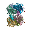
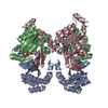
- 構造の表示
構造の表示
| 構造ビューア | 分子: MolmilJmol/JSmol |
|---|
- ダウンロードとリンク
-ダウンロード
| PDBx/mmCIF形式 | 1hnu.cif.gz | 68.1 KB | 表示 | PDBx/mmCIF形式 |
|---|---|---|---|---|
| PDB形式 | pdb1hnu.ent.gz | 50.6 KB | 表示 | PDB形式 |
| PDBx/mmJSON形式 | 1hnu.json.gz | ツリー表示 | PDBx/mmJSON形式 | |
| その他 |  その他のダウンロード その他のダウンロード |
-検証レポート
| アーカイブディレクトリ | https://data.pdbj.org/pub/pdb/validation_reports/hn/1hnuftp://data.pdbj.org/pub/pdb/validation_reports/hn/1hnu | HTTPS FTP |
|---|
-関連構造データ


-リンク
 PDBj
PDBj
- 集合体
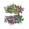
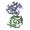
集合体
| 登録構造単位 | 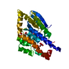
| |||||||||||||||
|---|---|---|---|---|---|---|---|---|---|---|---|---|---|---|---|---|
| 1 | x 6
| |||||||||||||||
| 2 | 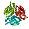
| |||||||||||||||
| 単位格子 |
| |||||||||||||||
| Components on special symmetry positions |
|
-要素
| #1: タンパク質 | 分子量: 31718.369 Da / 分子数: 1 / 由来タイプ: 組換発現 由来: (組換発現) プラスミド: PET3A / 発現宿主:  | ||||
|---|---|---|---|---|---|
| #2: 化合物 |   分子量: 250.205 Da / 分子数: 2 / 由来タイプ: 合成 / 式: O4Re 分子量: 250.205 Da / 分子数: 2 / 由来タイプ: 合成 / 式: O4Re#3: 化合物 |   分子量: 62.068 Da / 分子数: 2 / 由来タイプ: 合成 / 式: C2H6O2 分子量: 62.068 Da / 分子数: 2 / 由来タイプ: 合成 / 式: C2H6O2#4: 水 | ChemComp-HOH / |  分子量: 18.015 Da / 分子数: 194 / 由来タイプ: 天然 / 式: H2O 分子量: 18.015 Da / 分子数: 194 / 由来タイプ: 天然 / 式: H2O |
-実験情報
-実験
| 実験 | 手法: X線回折 / 使用した結晶の数: 1 |
|---|
- 試料調製
試料調製
| 結晶 | マシュー密度: 3.78 Å3/Da / 溶媒含有率: 67.48 % | ||||||||||||||||||||
|---|---|---|---|---|---|---|---|---|---|---|---|---|---|---|---|---|---|---|---|---|---|
| 結晶化 | 温度: 295 K / 手法: 蒸気拡散法, ハンギングドロップ法 / pH: 5.6 詳細: MES, ammonium sulfate, 1,4-dioxane, pH 5.6, VAPOR DIFFUSION, HANGING DROP, temperature 295K | ||||||||||||||||||||
| 結晶化 | *PLUS 手法: unknown | ||||||||||||||||||||
| 溶液の組成 | *PLUS
|
-データ収集
| 回折 | 平均測定温度: 100 K | |||||||||||||||
|---|---|---|---|---|---|---|---|---|---|---|---|---|---|---|---|---|
| 放射光源 | 由来: シンクロトロン / サイト: EMBL/DESY, HAMBURG  / ビームライン: BW7A / 波長: 1.1765, 1.1773, 1.1697, 1.1836 / ビームライン: BW7A / 波長: 1.1765, 1.1773, 1.1697, 1.1836 | |||||||||||||||
| 検出器 | タイプ: MARRESEARCH / 検出器: CCD / 日付: 1999年11月26日 | |||||||||||||||
| 放射 | モノクロメーター: synchrotron / プロトコル: MAD / 単色(M)・ラウエ(L): M / 散乱光タイプ: x-ray | |||||||||||||||
| 放射波長 |
| |||||||||||||||
| 反射 | 解像度: 2.15→30 Å / Num. all: 27191 / Num. obs: 27191 / % possible obs: 99.5 % / Observed criterion σ(F): 0 / Observed criterion σ(I): -3 / 冗長度: 4.8 % / Biso Wilson estimate: 40.97 Å2 / Rmerge(I) obs: 0.053 / Net I/σ(I): 16 | |||||||||||||||
| 反射 シェル | 解像度: 2.15→2.23 Å / 冗長度: 4.8 % / Rmerge(I) obs: 0.474 / Mean I/σ(I) obs: 2.9 / Num. unique all: 2651 / % possible all: 99.7 | |||||||||||||||
| 反射 | *PLUS 最低解像度: 30 Å | |||||||||||||||
| 反射 シェル | *PLUS % possible obs: 99.7 % / Num. unique obs: 2651 |
- 解析
解析
| ソフトウェア |
| ||||||||||||||||||||
|---|---|---|---|---|---|---|---|---|---|---|---|---|---|---|---|---|---|---|---|---|---|
| 精密化 | 構造決定の手法: 多波長異常分散 / 解像度: 2.15→20 Å / Isotropic thermal model: individual isotropic / σ(F): 0 / σ(I): 0 / 立体化学のターゲット値: Engh & Huber
| ||||||||||||||||||||
| 原子変位パラメータ | Biso mean: 44.95 Å2 | ||||||||||||||||||||
| 精密化ステップ | サイクル: LAST / 解像度: 2.15→20 Å
| ||||||||||||||||||||
| 拘束条件 |
| ||||||||||||||||||||
| LS精密化 シェル | 解像度: 2.15→2.25 Å /
| ||||||||||||||||||||
| ソフトウェア | *PLUS 名称: REFMAC / 分類: refinement | ||||||||||||||||||||
| 精密化 | *PLUS 最低解像度: 20 Å / σ(F): 0 | ||||||||||||||||||||
| 溶媒の処理 | *PLUS | ||||||||||||||||||||
| 原子変位パラメータ | *PLUS | ||||||||||||||||||||
| 拘束条件 | *PLUS
|
