- PDB-2i5i: CRYSTAL STRUCTURE OF A PUTATIVE CELLOBIOSE-PHOSPHATE CLEAVAGE PRO... -
+
Open data
ID or keywords:
Loading...
-
Basic information
Entry
Database: PDB / ID: 2i5i
Title
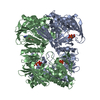
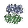
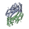
CRYSTAL STRUCTURE OF A PUTATIVE CELLOBIOSE-PHOSPHATE CLEAVAGE PROTEIN (EF3048) FROM ENTEROCOCCUS FAECALIS V583 AT 1.70 A RESOLUTION
Components
UPF0249 protein EF_3048
Keywords
HYDROLASE / PUTATIVE CELLOBIOSE-PHOSPHATE CLEAVAGE PROTEIN / STRUCTURAL GENOMICS / JOINT CENTER FOR STRUCTURAL GENOMICS / JCSG / PROTEIN STRUCTURE INITIATIVE / PSI-2
Function / homology
Function and homology information
hydrolase activity, acting on carbon-nitrogen (but not peptide) bonds, in linear amides / deacetylase activity / Hydrolases; Acting on carbon-nitrogen bonds, other than peptide bonds; In linear amides / polysaccharide catabolic process / metal ion binding Similarity search - Function
BIOMOLECULE: 1 THIS ENTRY CONTAINS THE CRYSTALLOGRAPHIC ASYMMETRIC UNIT WHICH CONSISTS OF 2 CHAIN(S) ...BIOMOLECULE: 1 THIS ENTRY CONTAINS THE CRYSTALLOGRAPHIC ASYMMETRIC UNIT WHICH CONSISTS OF 2 CHAIN(S). SEE REMARK 350 FOR INFORMATION ON GENERATING THE BIOLOGICAL MOLECULE(S). SIZE EXCLUSION CHROMATOGRAPHY SUPPORTS THE ASSIGNMENT OF A HEXAMER AS THE BIOLOGICALLY SIGNIFICANT OLIGIMERIZATION STATE.
Remark 999
SEQUENCE THE CONSTRUCT WAS EXPRESSED WITH A PURIFICATION TAG MGSDKIHHHHHHENLYFQG. THE TAG WAS ...SEQUENCE THE CONSTRUCT WAS EXPRESSED WITH A PURIFICATION TAG MGSDKIHHHHHHENLYFQG. THE TAG WAS REMOVED WITH TEV PROTEASE LEAVING ONLY A GLYCINE (0) FOLLOWED BY THE TARGET SEQUENCE.
Monochromator: Single crystal Si(111) bent monochromator (horizontal focusing) Protocol: MAD / Monochromatic (M) / Laue (L): M / Scattering type: x-ray
Radiation wavelength
ID
Wavelength (Å)
Relative weight
1
0.91837
1
2
0.978981
1
3
0.979291
1
Reflection
Resolution: 1.7→29.45 Å / Num. obs: 59414 / % possible obs: 99.6 % / Redundancy: 3.6 % / Rmerge(I) obs: 0.08 / Χ2: 1.016 / Net I/σ(I): 20.8
Reflection shell
Diffraction-ID: 1
Resolution (Å)
Redundancy (%)
Rmerge(I) obs
Mean I/σ(I) obs
Num. unique all
Χ2
% possible all
1.7-1.74
3.7
0.505
2
4243
1.02
100
1.74-1.79
3.7
0.408
4279
1.004
100
1.79-1.84
3.8
0.347
4225
0.948
100
1.84-1.9
3.7
0.286
4279
1.045
99.9
1.9-1.97
3.6
0.229
4262
1.015
99.7
1.97-2.05
3.7
0.178
4238
1.039
100
2.05-2.14
3.6
0.149
4241
1.048
99.5
2.14-2.25
3.5
0.124
4240
0.937
99.5
2.25-2.4
3.6
0.105
4242
1.124
99.9
2.4-2.58
3.5
0.093
4264
1.027
99.8
2.58-2.84
3.5
0.088
4260
1.029
99.7
2.84-3.25
3.4
0.075
4259
1.066
99.7
3.25-4.1
3.3
0.057
4201
1.035
98.9
4.1-50
3.5
0.052
4181
0.889
98.1
-
Phasing
Phasing
Method: MAD
-
Processing
Software
Name
Version
Classification
NB
MolProbity
3beta29
modelbuilding
SHELX
refinement
SCALEPACK
datascaling
PDB_EXTRACT
2
dataextraction
HKL-2000
datareduction
SOLVE
phasing
SHELXL-97
refinement
Refinement
Method to determine structure: MAD / Resolution: 1.7→29.45 Å / Num. parameters: 17818 / Num. restraintsaints: 22295 / Cross valid method: FREE R / σ(F): 0 / Stereochemistry target values: ENGH AND HUBER Details: OTHER REFINEMENT REMARKS: 1. HYDROGENS HAVE BEEN ADDED IN THE RIDING POSITIONS. 2. NCS RESTRAINTS WERE APPLIED BETWEEN RESIDUES 2-262 OF CHAINS A AND B. 3. REFINEMENT WAS AGAINST INTENSITY ...Details: OTHER REFINEMENT REMARKS: 1. HYDROGENS HAVE BEEN ADDED IN THE RIDING POSITIONS. 2. NCS RESTRAINTS WERE APPLIED BETWEEN RESIDUES 2-262 OF CHAINS A AND B. 3. REFINEMENT WAS AGAINST INTENSITY DATA. 4. REFINEMENT WAS CARRIED WITH THE TWIN LAW OF [K,H,-L] WITH THE TWIN FRACTION OF 0.3513. THE RFREE DECREASES FROM 0.27 TO 0.1888. 5. A MET-INHIBITION PROTOCOL WAS USED FOR SELENOMETHIONINE INCORPORATION DURING PROTEIN EXPRESSION. THE OCCUPANCY OF THE SE ATOMS IN THE MSE RESIDUES WAS REDUCED TO 0.75 TO ACCOUNT FOR THE REDUCED SCATTERING POWER DUE TO PARTIAL S-MET INCORPORATION.
In the structure databanks used in Yorodumi, some data are registered as the other names, "COVID-19 virus" and "2019-nCoV". Here are the details of the virus and the list of structure data.
Jan 31, 2019. EMDB accession codes are about to change! (news from PDBe EMDB page)
EMDB accession codes are about to change! (news from PDBe EMDB page)
The allocation of 4 digits for EMDB accession codes will soon come to an end. Whilst these codes will remain in use, new EMDB accession codes will include an additional digit and will expand incrementally as the available range of codes is exhausted. The current 4-digit format prefixed with “EMD-” (i.e. EMD-XXXX) will advance to a 5-digit format (i.e. EMD-XXXXX), and so on. It is currently estimated that the 4-digit codes will be depleted around Spring 2019, at which point the 5-digit format will come into force.
The EM Navigator/Yorodumi systems omit the EMD- prefix.
Related info.:Q: What is EMD? / ID/Accession-code notation in Yorodumi/EM Navigator
Yorodumi is a browser for structure data from EMDB, PDB, SASBDB, etc.
This page is also the successor to EM Navigator detail page, and also detail information page/front-end page for Omokage search.
The word "yorodu" (or yorozu) is an old Japanese word meaning "ten thousand". "mi" (miru) is to see.
Related info.:EMDB / PDB / SASBDB / Comparison of 3 databanks / Yorodumi Search / Aug 31, 2016. New EM Navigator & Yorodumi / Yorodumi Papers / Jmol/JSmol / Function and homology information / Changes in new EM Navigator and Yorodumi
 Movie
Movie Controller

 Yorodumi
 Open data
 Basic information
 Components
 Keywords
 Function and homology information
 Enterococcus faecalis (bacteria)
 X-RAY DIFFRACTION /
  Authors
 Citation
 Structure visualization
 Downloads & links
 Other downloads
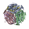


 PDBj
PDBj
 Assembly
 Mass: 18.015 Da / Num. of mol.: 363 / Source method: isolated from a natural source / Formula: H2O
 Sample preparation
 / Beamline: BL11-1 / Wavelength: 0.918370,0.978981,0.979291
 Processing