 Movie
Movie Controller

[English] 日本語
 Yorodumi
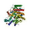
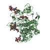
- PDB-2ze9: Crystal structure of H168A mutant of phospholipase D from Strepto... -
+ Open data

- Basic information
| Entry | Database: PDB / ID: 2ze9 | ||||||
|---|---|---|---|---|---|---|---|
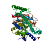
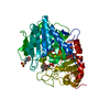
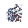
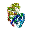
| Title | Crystal structure of H168A mutant of phospholipase D from Streptomyces antibioticus, as a complex with phosphatidylcholine | ||||||
 Components Components | Phospholipase D | ||||||
 Keywords Keywords | HYDROLASE / ALPHA-BETA-BETA-ALPHA SANDWICH / Lipid degradation / Secreted | ||||||
| Function / homology |  Function and homology information Function and homology informationphosphatidyltransferase activity / cardiolipin biosynthetic process / phospholipase D / D-type glycerophospholipase activity / lipid catabolic process / extracellular region Similarity search - Function | ||||||
| Biological species |  Streptomyces antibioticus (bacteria) Streptomyces antibioticus (bacteria) | ||||||
| Method |  X-RAY DIFFRACTION / MOLECULAR REPLACEMENT / Resolution: 2.3 Å X-RAY DIFFRACTION / MOLECULAR REPLACEMENT / Resolution: 2.3 Å | ||||||
 Authors Authors | Suzuki, A. / Toda, H. / Iwasaki, Y. / Yamane, T. / Yamane, T. | ||||||
 Citation Citation | Journal: To be Published Title: Crystal structure of phospholipase D from streptomyces antibioticus. Authors: Suzuki, A. / Toda, H. / Iwasaki, Y. / Yamane, T. / Yamane, T. #1: Journal: Acta Crystallogr.,Sect.D / Year: 1999 Title: Crystallization and preliminary X-ray diffraction studies of phospholipase D from Streptomyces antibioticus Authors: Suzuki, A. / Kakuno, K. / Iwasaki, Y. / Yamane, T. / Yamane, T. #2: Journal: Chembiochem / Year: 2008 Title: Streptomyces phospholipase D mutants with altered substrate specificity capable of phosphatidylinositol synthesis Authors: Masayama, A. / Takahashi, T. / Tsukada, K. / Nishikawa, S. / Takahashi, R. / Adachi, M. / Koga, K. / Suzuki, A. / Yamane, T. / Nakano, H. / Iwasaki, Y. | ||||||
| History |
|
- Structure visualization
| Structure viewer | Molecule: MolmilJmol/JSmol |
|---|
- Downloads & links
-Download
| PDBx/mmCIF format | 2ze9.cif.gz | 117.8 KB | Display | PDBx/mmCIF format |
|---|---|---|---|---|
| PDB format | pdb2ze9.ent.gz | 88.5 KB | Display | PDB format |
| PDBx/mmJSON format | 2ze9.json.gz | Tree view | PDBx/mmJSON format | |
| Others |  Other downloads Other downloads |
-Validation report
| Arichive directory | https://data.pdbj.org/pub/pdb/validation_reports/ze/2ze9ftp://data.pdbj.org/pub/pdb/validation_reports/ze/2ze9 | HTTPS FTP |
|---|
-Related structure data
| Related structure data |  2ze4S S: Starting model for refinement |
|---|---|
| Similar structure data |

-Links
 PDBj
PDBj- Assembly
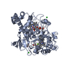
| Deposited unit | 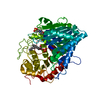
| ||||||||
|---|---|---|---|---|---|---|---|---|---|
| 1 |
| ||||||||
| Unit cell |
|
-Components
| #1: Protein | Mass: 53968.992 Da / Num. of mol.: 1 / Mutation: H168A Source method: isolated from a genetically manipulated source Source: (gene. exp.) Streptomyces antibioticus (bacteria) / Plasmid: pPELB-PLD3 / Species (production host): Escherichia coli / Production host: |
|---|---|
| #2: Chemical | ChemComp-MES /   Mass: 195.237 Da / Num. of mol.: 1 / Source method: obtained synthetically / Formula: C6H13NO4S / Comment: pH buffer*YM Mass: 195.237 Da / Num. of mol.: 1 / Source method: obtained synthetically / Formula: C6H13NO4S / Comment: pH buffer*YM |
| #3: Chemical | ChemComp-PD7 / (  Mass: 396.413 Da / Num. of mol.: 1 / Source method: obtained synthetically / Formula: C17H33O8P Mass: 396.413 Da / Num. of mol.: 1 / Source method: obtained synthetically / Formula: C17H33O8P |
| #4: Water | ChemComp-HOH /  Mass: 18.015 Da / Num. of mol.: 365 / Source method: isolated from a natural source / Formula: H2O Mass: 18.015 Da / Num. of mol.: 365 / Source method: isolated from a natural source / Formula: H2O |
| Has protein modification | Y |
| Sequence details | THERE ARE DIFFERENCES BETWEEN THE SEQRES AND THE SEQUENCE DATABASE. ACCORDING TO THE DEPOSITOR, THE ...THERE ARE DIFFERENCE |
-Experimental details
-Experiment
| Experiment | Method: X-RAY DIFFRACTION / Number of used crystals: 1 |
|---|
- Sample preparation
| Crystal | Density Matthews: 2.17 Å3/Da / Density % sol: 43.45 % |
|---|---|
| Crystal grow | Temperature: 291 K / Method: vapor diffusion, hanging drop / pH: 6 Details: 15% PEG 6000, 0.1M MES, pH 6.0, VAPOR DIFFUSION, HANGING DROP, temperature 291K |
-Data collection
| Diffraction | Mean temperature: 100 K |
|---|---|
| Diffraction source | Source: ROTATING ANODE / Type: RIGAKU RU300 / Wavelength: 1.5418 Å |
| Detector | Type: RIGAKU RAXIS IV / Detector: IMAGE PLATE / Date: Jul 1, 1999 / Details: mirrors |
| Radiation | Monochromator: YALE MIRRORS / Protocol: SINGLE WAVELENGTH / Monochromatic (M) / Laue (L): M / Scattering type: x-ray |
| Radiation wavelength | Wavelength: 1.5418 Å / Relative weight: 1 |
| Reflection | Resolution: 2.3→61.31 Å / Num. all: 21448 / Num. obs: 20805 / % possible obs: 97 % / Observed criterion σ(F): 0 / Observed criterion σ(I): 0 / Redundancy: 3.4 % / Biso Wilson estimate: 23.6 Å2 / Rmerge(I) obs: 0.07 / Net I/σ(I): 17.2 |
| Reflection shell | Resolution: 2.3→2.42 Å / Redundancy: 2.2 % / Rmerge(I) obs: 0.157 / Mean I/σ(I) obs: 5.5 / Num. unique all: 2726 / % possible all: 88.8 |
- Processing
| Software |
| ||||||||||||||||||||||||||||||||||||||||||||||||||||||||||||||||||||||||||||||||||||||||||
|---|---|---|---|---|---|---|---|---|---|---|---|---|---|---|---|---|---|---|---|---|---|---|---|---|---|---|---|---|---|---|---|---|---|---|---|---|---|---|---|---|---|---|---|---|---|---|---|---|---|---|---|---|---|---|---|---|---|---|---|---|---|---|---|---|---|---|---|---|---|---|---|---|---|---|---|---|---|---|---|---|---|---|---|---|---|---|---|---|---|---|---|
| Refinement | Method to determine structure: MOLECULAR REPLACEMENT Starting model: PDB ENTRY 2ZE4 Resolution: 2.3→48.04 Å / Cor.coef. Fo:Fc: 0.937 / Cor.coef. Fo:Fc free: 0.879 / SU B: 6.9 / SU ML: 0.17 / Isotropic thermal model: Isotropic / Cross valid method: THROUGHOUT / σ(F): 0 / ESU R: 0.481 / ESU R Free: 0.275 / Stereochemistry target values: MAXIMUM LIKELIHOOD / Details: HYDROGENS HAVE BEEN ADDED IN THE RIDING POSITIONS
| ||||||||||||||||||||||||||||||||||||||||||||||||||||||||||||||||||||||||||||||||||||||||||
| Solvent computation | Ion probe radii: 0.8 Å / Shrinkage radii: 0.8 Å / VDW probe radii: 1.2 Å / Solvent model: MASK | ||||||||||||||||||||||||||||||||||||||||||||||||||||||||||||||||||||||||||||||||||||||||||
| Displacement parameters | Biso mean: 13.618 Å2
| ||||||||||||||||||||||||||||||||||||||||||||||||||||||||||||||||||||||||||||||||||||||||||
| Refinement step | Cycle: LAST / Resolution: 2.3→48.04 Å
| ||||||||||||||||||||||||||||||||||||||||||||||||||||||||||||||||||||||||||||||||||||||||||
| Refine LS restraints |
| ||||||||||||||||||||||||||||||||||||||||||||||||||||||||||||||||||||||||||||||||||||||||||
| LS refinement shell | Resolution: 2.3→2.359 Å / Total num. of bins used: 20
|
