Type: MARMOSAIC 225 mm CCD / Detector: CCD / Date: Jun 15, 2007 Details: A fixed exit Si double crystal monochromator followed by a two dimensional focusing mirror which is coated in rhodium.
Radiation
Monochromator: Fixed exit Si double crystal monochromator / Protocol: SINGLE WAVELENGTH / Monochromatic (M) / Laue (L): M / Scattering type: x-ray
Radiation wavelength
Wavelength: 1 Å / Relative weight: 1
Reflection
Resolution: 1.9→50 Å / Num. all: 115364 / Num. obs: 114964 / % possible obs: 99.8 % / Redundancy: 6.9 % / Biso Wilson estimate: 14.2 Å2 / Rmerge(I) obs: 0.081 / Rsym value: 0.078 / Net I/σ(I): 23.24
Reflection shell
Resolution: 1.9→1.97 Å / Redundancy: 5.4 % / Rmerge(I) obs: 0.354 / Mean I/σ(I) obs: 4.09 / Num. unique all: 11187 / Rsym value: 0.317 / % possible all: 97.9
-
Processing
Software
Name
Version
Classification
CNS
1.1
refinement
BSS
datacollection
HKL-2000
datareduction
HKL-2000
datascaling
MOLREP
phasing
Refinement
Method to determine structure: MOLECULAR REPLACEMENT Starting model: 2ZDG
In the structure databanks used in Yorodumi, some data are registered as the other names, "COVID-19 virus" and "2019-nCoV". Here are the details of the virus and the list of structure data.
Jan 31, 2019. EMDB accession codes are about to change! (news from PDBe EMDB page)
EMDB accession codes are about to change! (news from PDBe EMDB page)
The allocation of 4 digits for EMDB accession codes will soon come to an end. Whilst these codes will remain in use, new EMDB accession codes will include an additional digit and will expand incrementally as the available range of codes is exhausted. The current 4-digit format prefixed with “EMD-” (i.e. EMD-XXXX) will advance to a 5-digit format (i.e. EMD-XXXXX), and so on. It is currently estimated that the 4-digit codes will be depleted around Spring 2019, at which point the 5-digit format will come into force.
The EM Navigator/Yorodumi systems omit the EMD- prefix.
Related info.:Q: What is EMD? / ID/Accession-code notation in Yorodumi/EM Navigator
Yorodumi is a browser for structure data from EMDB, PDB, SASBDB, etc.
This page is also the successor to EM Navigator detail page, and also detail information page/front-end page for Omokage search.
The word "yorodu" (or yorozu) is an old Japanese word meaning "ten thousand". "mi" (miru) is to see.
Related info.:EMDB / PDB / SASBDB / Comparison of 3 databanks / Yorodumi Search / Aug 31, 2016. New EM Navigator & Yorodumi / Yorodumi Papers / Jmol/JSmol / Function and homology information / Changes in new EM Navigator and Yorodumi
 Movie
Movie Controller

 Yorodumi
 Open data
 Basic information
 Components
 Keywords
 Function and homology information
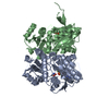
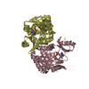
 Thermus thermophilus (bacteria)
 X-RAY DIFFRACTION /
  Authors
 Citation
 Structure visualization
 Downloads & links
 Other downloads


 PDBj
PDBj


 Assembly

 Mass: 24.305 Da / Num. of mol.: 7 / Source method: obtained synthetically / Formula: Mg
 Mass: 427.201 Da / Num. of mol.: 4 / Source method: obtained synthetically / Formula: C10H15N5O10P2 / Comment: ADP, energy-carrying molecule*YM
 Type: D-peptide linking / Mass: 89.093 Da / Num. of mol.: 2 / Source method: obtained synthetically / Formula: C3H7NO2
 Mass: 18.015 Da / Num. of mol.: 643 / Source method: isolated from a natural source / Formula: H2O
 Sample preparation
 / Beamline: BL26B2 / Wavelength: 1 Å
 Processing