 Movie
Movie Controller

[English] 日本語
 Yorodumi
- PDB-2zb7: Crystal structure of human 15-ketoprostaglandin delta-13-reductas... -
+ Open data

- Basic information
| Entry | Database: PDB / ID: 2zb7 | ||||||
|---|---|---|---|---|---|---|---|
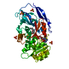
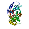
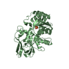
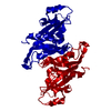
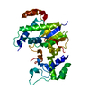
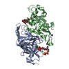
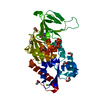
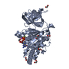
| Title | Crystal structure of human 15-ketoprostaglandin delta-13-reductase in complex with NADPH and nicotinamide | ||||||
 Components Components | Prostaglandin reductase 2 | ||||||
 Keywords Keywords | OXIDOREDUCTASE / Rossmann fold / Alternative splicing / Cytoplasm / NADP | ||||||
| Function / homology |  Function and homology information Function and homology information13,14-dehydro-15-oxoprostaglandin 13-reductase / 15-oxoprostaglandin 13-reductase [NAD(P)+] activity / Synthesis of Prostaglandins (PG) and Thromboxanes (TX) / prostaglandin metabolic process / cytoplasm Similarity search - Function | ||||||
| Biological species |  Homo sapiens (human) Homo sapiens (human) | ||||||
| Method |  X-RAY DIFFRACTION / MOLECULAR REPLACEMENT / Resolution: 1.8 Å X-RAY DIFFRACTION / MOLECULAR REPLACEMENT / Resolution: 1.8 Å | ||||||
 Authors Authors | Wu, Y.H. / Wang, A.H.J. / Ko, T.P. / Guo, R.T. / Hu, S.M. / Chuang, L.M. | ||||||
 Citation Citation | Journal: Structure / Year: 2008 Title: Structural basis for catalytic and inhibitory mechanisms of human prostaglandin reductase PTGR2. Authors: Wu, Y.H. / Ko, T.P. / Guo, R.T. / Hu, S.M. / Chuang, L.M. / Wang, A.H.J. | ||||||
| History |
|
- Structure visualization
| Structure viewer | Molecule: MolmilJmol/JSmol |
|---|
- Downloads & links
-Download
| PDBx/mmCIF format | 2zb7.cif.gz | 93 KB | Display | PDBx/mmCIF format |
|---|---|---|---|---|
| PDB format | pdb2zb7.ent.gz | 69.3 KB | Display | PDB format |
| PDBx/mmJSON format | 2zb7.json.gz | Tree view | PDBx/mmJSON format | |
| Others |  Other downloads Other downloads |
-Validation report
| Arichive directory | https://data.pdbj.org/pub/pdb/validation_reports/zb/2zb7ftp://data.pdbj.org/pub/pdb/validation_reports/zb/2zb7 | HTTPS FTP |
|---|
-Related structure data
| Related structure data | 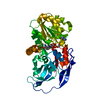 2zb3SC  2zb4C 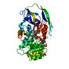 2zb8C S: Starting model for refinement C: citing same article ( |
|---|---|
| Similar structure data |


-Links
 PDBj
PDBj


- Assembly
| Deposited unit | 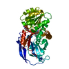
| ||||||||
|---|---|---|---|---|---|---|---|---|---|
| 1 |
| ||||||||
| Unit cell |
|
-Components
| #1: Protein | Mass: 38961.285 Da / Num. of mol.: 1 Source method: isolated from a genetically manipulated source Source: (gene. exp.) Homo sapiens (human) / Gene: PTGR2, ZADH1 / Plasmid: pET32 / Production host:  References: UniProt: Q8N8N7, 13,14-dehydro-15-oxoprostaglandin 13-reductase |
|---|---|
| #2: Chemical | ChemComp-NDP /   Mass: 745.421 Da / Num. of mol.: 1 / Source method: obtained synthetically / Formula: C21H30N7O17P3 Mass: 745.421 Da / Num. of mol.: 1 / Source method: obtained synthetically / Formula: C21H30N7O17P3 |
| #3: Chemical | ChemComp-NCA /   Mass: 122.125 Da / Num. of mol.: 1 / Source method: obtained synthetically / Formula: C6H6N2O / Comment: medication*YM Mass: 122.125 Da / Num. of mol.: 1 / Source method: obtained synthetically / Formula: C6H6N2O / Comment: medication*YM |
| #4: Water | ChemComp-HOH /  Mass: 18.015 Da / Num. of mol.: 446 / Source method: isolated from a natural source / Formula: H2O Mass: 18.015 Da / Num. of mol.: 446 / Source method: isolated from a natural source / Formula: H2O |
-Experimental details
-Experiment
| Experiment | Method: X-RAY DIFFRACTION / Number of used crystals: 1 |
|---|
- Sample preparation
| Crystal | Density Matthews: 2.14 Å3/Da / Density % sol: 42.49 % |
|---|---|
| Crystal grow | Temperature: 298 K / Method: vapor diffusion, hanging drop / pH: 5.9 Details: 0.1M MES, 2.0M ammonium sulfate, 2mM DTT, pH 5.9, VAPOR DIFFUSION, HANGING DROP, temperature 298K |
-Data collection
| Diffraction | Mean temperature: 100 K |
|---|---|
| Diffraction source | Source: ROTATING ANODE / Type: RIGAKU MICROMAX-007 HF / Wavelength: 1.5418 Å |
| Detector | Type: RIGAKU RAXIS IV++ / Detector: IMAGE PLATE / Date: Nov 16, 2005 / Details: MSC Varimax confocal |
| Radiation | Monochromator: confocal optical mirror / Protocol: SINGLE WAVELENGTH / Monochromatic (M) / Laue (L): M / Scattering type: x-ray |
| Radiation wavelength | Wavelength: 1.5418 Å / Relative weight: 1 |
| Reflection | Resolution: 1.8→50 Å / Num. all: 31865 / Num. obs: 28551 / % possible obs: 89.6 % / Observed criterion σ(F): 0 / Observed criterion σ(I): 1 / Redundancy: 5.9 % / Rmerge(I) obs: 0.051 / Net I/σ(I): 51.8 |
| Reflection shell | Resolution: 1.8→1.86 Å / Redundancy: 3.7 % / Rmerge(I) obs: 0.348 / Mean I/σ(I) obs: 6.4 / Num. unique all: 2065 / % possible all: 66.4 |
- Processing
| Software |
| |||||||||||||||||||||||||
|---|---|---|---|---|---|---|---|---|---|---|---|---|---|---|---|---|---|---|---|---|---|---|---|---|---|---|
| Refinement | Method to determine structure: MOLECULAR REPLACEMENT Starting model: PDB entry 2ZB3 Resolution: 1.8→50 Å / Isotropic thermal model: Isotropic / Cross valid method: THROUGHOUT / σ(F): 0 / σ(I): 0 / Stereochemistry target values: Engh & Huber
| |||||||||||||||||||||||||
| Displacement parameters | Biso mean: 23 Å2 | |||||||||||||||||||||||||
| Refine analyze |
| |||||||||||||||||||||||||
| Refinement step | Cycle: LAST / Resolution: 1.8→50 Å
| |||||||||||||||||||||||||
| Refine LS restraints |
| |||||||||||||||||||||||||
| LS refinement shell | Resolution: 1.8→1.86 Å / Rfactor Rfree error: 0.028
|
