 Movie
Movie Controller

[English] 日本語
 Yorodumi
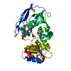
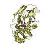
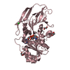
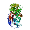
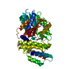
- PDB-3h8w: Structure of D132N T4 RNase H in the presence of divalent magnesium -
+ Open data

- Basic information
| Entry | Database: PDB / ID: 3h8w | ||||||
|---|---|---|---|---|---|---|---|
| Title | Structure of D132N T4 RNase H in the presence of divalent magnesium | ||||||
 Components Components | Ribonuclease H | ||||||
 Keywords Keywords | HYDROLASE / BPT4 RNase H / 5'-3' exonuclease / Endonuclease / Nuclease | ||||||
| Function / homology |  Function and homology information Function and homology informationDNA replication, Okazaki fragment processing / 5'-3' RNA exonuclease activity / 5'-flap endonuclease activity / DNA replication, removal of RNA primer / ribonuclease H / RNA-DNA hybrid ribonuclease activity / DNA binding Similarity search - Function | ||||||
| Biological species |  Enterobacteria phage T4 (virus) Enterobacteria phage T4 (virus) | ||||||
| Method |  X-RAY DIFFRACTION / SYNCHROTRON / MOLECULAR REPLACEMENT / molecular replacement / Resolution: 2.8 Å X-RAY DIFFRACTION / SYNCHROTRON / MOLECULAR REPLACEMENT / molecular replacement / Resolution: 2.8 Å | ||||||
 Authors Authors | Tomanicek, S.J. / Mueser, T.C. | ||||||
 Citation Citation | Journal: TO BE PUBLISHED Title: Additional Order Appears in the Absence of Metals in a FEN-1 protein: Structural Analysis of Magnesium Binding to Bacteriophage T4 RNaseH Authors: Tomanicek, S.J. / Devos, J.M. / Mueser, T.C. | ||||||
| History |
|
- Structure visualization
| Structure viewer | Molecule: MolmilJmol/JSmol |
|---|
- Downloads & links
-Download
| PDBx/mmCIF format | 3h8w.cif.gz | 69.5 KB | Display | PDBx/mmCIF format |
|---|---|---|---|---|
| PDB format | pdb3h8w.ent.gz | 50.6 KB | Display | PDB format |
| PDBx/mmJSON format | 3h8w.json.gz | Tree view | PDBx/mmJSON format | |
| Others |  Other downloads Other downloads |
-Validation report
| Arichive directory | https://data.pdbj.org/pub/pdb/validation_reports/h8/3h8wftp://data.pdbj.org/pub/pdb/validation_reports/h8/3h8w | HTTPS FTP |
|---|
-Related structure data
| Related structure data |  3h7iC 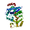 3h8jC 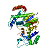 3h8sC 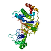 1tfrS S: Starting model for refinement C: citing same article ( |
|---|---|
| Similar structure data |


-Links
 PDBj
PDBj


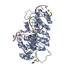
- Assembly
| Deposited unit | 
| ||||||||
|---|---|---|---|---|---|---|---|---|---|
| 1 |
| ||||||||
| Unit cell |
|
-Components
| #1: Protein | Mass: 35609.926 Da / Num. of mol.: 1 / Mutation: D132N Source method: isolated from a genetically manipulated source Source: (gene. exp.) Enterobacteria phage T4 (virus) / Strain: T4D / Gene: 33.2, das, rnh / Plasmid: pNN2202-D132N / Production host:  |
|---|---|
| #2: Water | ChemComp-HOH /  Mass: 18.015 Da / Num. of mol.: 27 / Source method: isolated from a natural source / Formula: H2O Mass: 18.015 Da / Num. of mol.: 27 / Source method: isolated from a natural source / Formula: H2O |
-Experimental details
-Experiment
| Experiment | Method: X-RAY DIFFRACTION / Number of used crystals: 1 |
|---|
- Sample preparation
| Crystal | Density Matthews: 2.48 Å3/Da / Density % sol: 50.35 % |
|---|---|
| Crystal grow | Temperature: 295 K / Method: vapor diffusion, hanging drop / pH: 6.5 Details: 100 mM Na PIPES, 200 mM MgCl2, 14% PEG 8000, pH 6.5, VAPOR DIFFUSION, HANGING DROP, temperature 295K |
-Data collection
| Diffraction | Mean temperature: 100 K |
|---|---|
| Diffraction source | Source: SYNCHROTRON / Site: APS  / Beamline: 14-BM-C / Wavelength: 1 Å / Beamline: 14-BM-C / Wavelength: 1 Å |
| Detector | Type: ADSC QUANTUM 4 / Detector: CCD |
| Radiation | Protocol: SINGLE WAVELENGTH / Monochromatic (M) / Laue (L): M / Scattering type: x-ray |
| Radiation wavelength | Wavelength: 1 Å / Relative weight: 1 |
| Reflection | Resolution: 2.8→34.34 Å / Num. obs: 8890 / % possible obs: 97.4 % / Redundancy: 3.6 % / Rmerge(I) obs: 0.041 / Rsym value: 0.041 / Net I/σ(I): 15.3 |
| Reflection shell | Resolution: 2.8→2.95 Å / Redundancy: 3.4 % / Rmerge(I) obs: 0.177 / Mean I/σ(I) obs: 4.3 / Num. measured all: 4280 / Num. unique all: 1242 / Rsym value: 0.177 / % possible all: 94.5 |
-Phasing
| Phasing | Method: molecular replacement |
|---|
- Processing
| Software |
| |||||||||||||||||||||||||||||||||||||||||||||||||
|---|---|---|---|---|---|---|---|---|---|---|---|---|---|---|---|---|---|---|---|---|---|---|---|---|---|---|---|---|---|---|---|---|---|---|---|---|---|---|---|---|---|---|---|---|---|---|---|---|---|---|
| Refinement | Method to determine structure: MOLECULAR REPLACEMENT Starting model: pdb entry 1TFR native T4 RNase H Resolution: 2.8→34.34 Å / Occupancy max: 1 / Occupancy min: 1 / SU ML: 0.48 / σ(F): 1.35 / Stereochemistry target values: ML
| |||||||||||||||||||||||||||||||||||||||||||||||||
| Solvent computation | Shrinkage radii: 0.9 Å / VDW probe radii: 1.11 Å / Solvent model: FLAT BULK SOLVENT MODEL / Bsol: 35.949 Å2 / ksol: 0.315 e/Å3 | |||||||||||||||||||||||||||||||||||||||||||||||||
| Displacement parameters | Biso max: 70.25 Å2 / Biso mean: 44.94 Å2 / Biso min: 48.45 Å2
| |||||||||||||||||||||||||||||||||||||||||||||||||
| Refinement step | Cycle: LAST / Resolution: 2.8→34.34 Å
| |||||||||||||||||||||||||||||||||||||||||||||||||
| Refine LS restraints |
| |||||||||||||||||||||||||||||||||||||||||||||||||
| LS refinement shell | Refine-ID: X-RAY DIFFRACTION / Total num. of bins used: 6
|
