 Movie
Movie Controller

[English] 日本語
 Yorodumi
- PDB-2zb3: Crystal structure of mouse 15-ketoprostaglandin delta-13-reductas... -
+ Open data

- Basic information
| Entry | Database: PDB / ID: 2zb3 | ||||||
|---|---|---|---|---|---|---|---|
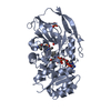
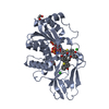
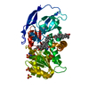
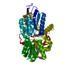
| Title | Crystal structure of mouse 15-ketoprostaglandin delta-13-reductase in complex with NADPH | ||||||
 Components Components | Prostaglandin reductase 2 | ||||||
 Keywords Keywords | OXIDOREDUCTASE / Rossmann fold | ||||||
| Function / homology |  Function and homology information Function and homology information13,14-dehydro-15-oxoprostaglandin 13-reductase / 15-oxoprostaglandin 13-reductase [NAD(P)+] activity / prostaglandin metabolic process / mitochondrion / cytosol Similarity search - Function | ||||||
| Biological species |  | ||||||
| Method |  X-RAY DIFFRACTION / SYNCHROTRON / MOLECULAR REPLACEMENT / Resolution: 2 Å X-RAY DIFFRACTION / SYNCHROTRON / MOLECULAR REPLACEMENT / Resolution: 2 Å | ||||||
 Authors Authors | Wu, Y.H. / Wang, A.H.J. / Ko, T.P. / Guo, R.T. / Hu, S.M. / Chuang, L.M. | ||||||
 Citation Citation | Journal: Structure / Year: 2008 Title: Structural basis for catalytic and inhibitory mechanisms of human prostaglandin reductase PTGR2. Authors: Wu, Y.H. / Ko, T.P. / Guo, R.T. / Hu, S.M. / Chuang, L.M. / Wang, A.H.J. | ||||||
| History |
|
- Structure visualization
| Structure viewer | Molecule: MolmilJmol/JSmol |
|---|
- Downloads & links
-Download
| PDBx/mmCIF format | 2zb3.cif.gz | 92.8 KB | Display | PDBx/mmCIF format |
|---|---|---|---|---|
| PDB format | pdb2zb3.ent.gz | 69.2 KB | Display | PDB format |
| PDBx/mmJSON format | 2zb3.json.gz | Tree view | PDBx/mmJSON format | |
| Others |  Other downloads Other downloads |
-Validation report
| Arichive directory | https://data.pdbj.org/pub/pdb/validation_reports/zb/2zb3ftp://data.pdbj.org/pub/pdb/validation_reports/zb/2zb3 | HTTPS FTP |
|---|
-Related structure data
| Related structure data |  2zb4C 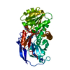 2zb7C 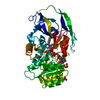 2zb8C 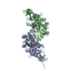 1v3vS C: citing same article ( S: Starting model for refinement |
|---|---|
| Similar structure data |

-Links
 PDBj
PDBj


- Assembly
| Deposited unit | 
| ||||||||
|---|---|---|---|---|---|---|---|---|---|
| 1 |
| ||||||||
| 2 | 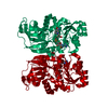
| ||||||||
| Unit cell |
| ||||||||
| Components on special symmetry positions |
|
-Components
| #1: Protein | Mass: 38198.285 Da / Num. of mol.: 1 Source method: isolated from a genetically manipulated source Source: (gene. exp.)  References: UniProt: Q8VDQ1, 13,14-dehydro-15-oxoprostaglandin 13-reductase |
|---|---|
| #2: Chemical | ChemComp-NDP /   Mass: 745.421 Da / Num. of mol.: 1 / Source method: obtained synthetically / Formula: C21H30N7O17P3 Mass: 745.421 Da / Num. of mol.: 1 / Source method: obtained synthetically / Formula: C21H30N7O17P3 |
| #3: Water | ChemComp-HOH /  Mass: 18.015 Da / Num. of mol.: 464 / Source method: isolated from a natural source / Formula: H2O Mass: 18.015 Da / Num. of mol.: 464 / Source method: isolated from a natural source / Formula: H2O |
| Sequence details | THE FEATURE OF UNIPROT (PTGR2_MOUSE, Q8VDQ1) SHOWS CONFLICT AT THIS POSITION: P -> T (IN REF. 1; BAB32284) |
-Experimental details
-Experiment
| Experiment | Method: X-RAY DIFFRACTION / Number of used crystals: 1 |
|---|
- Sample preparation
| Crystal | Density Matthews: 2.43 Å3/Da / Density % sol: 49.43 % |
|---|---|
| Crystal grow | Temperature: 298 K / Method: vapor diffusion, sitting drop / pH: 6.5 Details: 0.1M cacodylate, pH6.5, 27% PEG 8000, 80mM Mg(OAc)2, VAPOR DIFFUSION, SITTING DROP, temperature 298K |
-Data collection
| Diffraction | Mean temperature: 100 K |
|---|---|
| Diffraction source | Source: SYNCHROTRON / Site: SPring-8  / Beamline: BL12B2 / Wavelength: 1 Å / Beamline: BL12B2 / Wavelength: 1 Å |
| Detector | Type: ADSC QUANTUM 4 / Detector: CCD / Date: Nov 13, 2004 / Details: mirrors |
| Radiation | Monochromator: Si(111) / Protocol: SINGLE WAVELENGTH / Monochromatic (M) / Laue (L): M / Scattering type: x-ray |
| Radiation wavelength | Wavelength: 1 Å / Relative weight: 1 |
| Reflection | Resolution: 2→30 Å / Num. all: 26149 / Num. obs: 24685 / % possible obs: 94.4 % / Observed criterion σ(F): 0 / Observed criterion σ(I): 1 / Redundancy: 7.3 % / Rmerge(I) obs: 0.092 / Net I/σ(I): 19.4 |
| Reflection shell | Resolution: 2→2.07 Å / Redundancy: 5.9 % / Rmerge(I) obs: 0.969 / Mean I/σ(I) obs: 2.1 / Num. unique all: 2447 / % possible all: 96.1 |
- Processing
| Software |
| |||||||||||||||||||||||||
|---|---|---|---|---|---|---|---|---|---|---|---|---|---|---|---|---|---|---|---|---|---|---|---|---|---|---|
| Refinement | Method to determine structure: MOLECULAR REPLACEMENT Starting model: PDB ENTRY 1V3V Resolution: 2→30 Å / Isotropic thermal model: Isotropic / Cross valid method: THROUGHOUT / σ(F): 0 / σ(I): 0 / Stereochemistry target values: Engh & Huber
| |||||||||||||||||||||||||
| Displacement parameters | Biso mean: 33.2 Å2 | |||||||||||||||||||||||||
| Refine analyze |
| |||||||||||||||||||||||||
| Refinement step | Cycle: LAST / Resolution: 2→30 Å
| |||||||||||||||||||||||||
| Refine LS restraints |
| |||||||||||||||||||||||||
| LS refinement shell | Resolution: 2→2.07 Å / Rfactor Rfree error: 0.008
|
