 Movie
Movie Controller

[English] 日本語
 Yorodumi
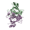
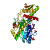
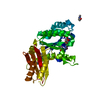
- PDB-1nh1: Crystal Structure of the Type III Effector AvrB from Pseudomonas ... -
+ Open data

- Basic information
| Entry | Database: PDB / ID: 1nh1 | ||||||
|---|---|---|---|---|---|---|---|
| Title | Crystal Structure of the Type III Effector AvrB from Pseudomonas syringae. | ||||||
 Components Components | Avirulence B protein | ||||||
 Keywords Keywords | Avirulence Protein / HELIX BUNDLE | ||||||
| Function / homology |  Function and homology information Function and homology information | ||||||
| Biological species |  Pseudomonas syringae (bacteria) Pseudomonas syringae (bacteria) | ||||||
| Method |  X-RAY DIFFRACTION / SYNCHROTRON / MAD / Resolution: 2.2 Å X-RAY DIFFRACTION / SYNCHROTRON / MAD / Resolution: 2.2 Å | ||||||
 Authors Authors | Lee, C.C. / Wood, M.D. / Ng, K. / Luginbuhl, P. / Spraggon, G. / Katagiri, F. | ||||||
 Citation Citation | Journal: Structure / Year: 2004 Title: Crystal Structure of the Type III Effector AvrB from Pseudomonas syringae. Authors: Lee, C.C. / Wood, M.D. / Ng, K. / Luginbuhl, P. / Spraggon, G. / Katagiri, F. | ||||||
| History |
|
- Structure visualization
| Structure viewer | Molecule: MolmilJmol/JSmol |
|---|
- Downloads & links
-Download
| PDBx/mmCIF format | 1nh1.cif.gz | 70.8 KB | Display | PDBx/mmCIF format |
|---|---|---|---|---|
| PDB format | pdb1nh1.ent.gz | 53.1 KB | Display | PDB format |
| PDBx/mmJSON format | 1nh1.json.gz | Tree view | PDBx/mmJSON format | |
| Others |  Other downloads Other downloads |
-Validation report
| Arichive directory | https://data.pdbj.org/pub/pdb/validation_reports/nh/1nh1ftp://data.pdbj.org/pub/pdb/validation_reports/nh/1nh1 | HTTPS FTP |
|---|
-Related structure data
| Similar structure data |
|---|


-Links
 PDBj
PDBj- Assembly
| Deposited unit | 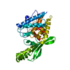
| ||||||||
|---|---|---|---|---|---|---|---|---|---|
| 1 |
| ||||||||
| Unit cell |
|
-Components
| #1: Protein | Mass: 37097.211 Da / Num. of mol.: 1 Source method: isolated from a genetically manipulated source Source: (gene. exp.) Pseudomonas syringae (bacteria) / Gene: AVRB / Plasmid: pET-27b(+) / Cell line (production host): BL21 (DE3) pLysS / Production host: |
|---|---|
| #2: Water | ChemComp-HOH /  Mass: 18.015 Da / Num. of mol.: 151 / Source method: isolated from a natural source / Formula: H2O Mass: 18.015 Da / Num. of mol.: 151 / Source method: isolated from a natural source / Formula: H2O |
-Experimental details
-Experiment
| Experiment | Method: X-RAY DIFFRACTION / Number of used crystals: 1 |
|---|
- Sample preparation
| Crystal | Density Matthews: 3.85 Å3/Da / Density % sol: 68.01 % | |||||||||||||||||||||||||||||||||||||||||||||||||
|---|---|---|---|---|---|---|---|---|---|---|---|---|---|---|---|---|---|---|---|---|---|---|---|---|---|---|---|---|---|---|---|---|---|---|---|---|---|---|---|---|---|---|---|---|---|---|---|---|---|---|
| Crystal grow | Method: vapor diffusion, hanging drop / Details: VAPOR DIFFUSION, HANGING DROP | |||||||||||||||||||||||||||||||||||||||||||||||||
| Crystal grow | *PLUS Temperature: 4 ℃ / pH: 7.9 / Method: vapor diffusion, hanging drop | |||||||||||||||||||||||||||||||||||||||||||||||||
| Components of the solutions | *PLUS
|
-Data collection
| Diffraction |
| |||||||||||||||
|---|---|---|---|---|---|---|---|---|---|---|---|---|---|---|---|---|
| Diffraction source |
| |||||||||||||||
| Detector |
| |||||||||||||||
| Radiation | Protocol: MAD / Monochromatic (M) / Laue (L): M / Scattering type: x-ray | |||||||||||||||
| Radiation wavelength | Relative weight: 1 | |||||||||||||||
| Reflection | Resolution: 2.2→50 Å / Num. all: 29594 / Num. obs: 27892 / % possible obs: 94.2 % | |||||||||||||||
| Reflection | *PLUS Lowest resolution: 50 Å / % possible obs: 93.4 % / Rmerge(I) obs: 0.066 | |||||||||||||||
| Reflection shell | *PLUS % possible obs: 64.6 % / Rmerge(I) obs: 0.493 |

- Processing
| Software | Name: CNS / Classification: refinement | ||||||||||||||||||||
|---|---|---|---|---|---|---|---|---|---|---|---|---|---|---|---|---|---|---|---|---|---|
| Refinement | Method to determine structure: MAD / Resolution: 2.2→50 Å
| ||||||||||||||||||||
| Refinement step | Cycle: LAST / Resolution: 2.2→50 Å
| ||||||||||||||||||||
| Refinement | *PLUS Lowest resolution: 500 Å / Rfactor Rfree: 0.275 | ||||||||||||||||||||
| Solvent computation | *PLUS | ||||||||||||||||||||
| Displacement parameters | *PLUS | ||||||||||||||||||||
| Refine LS restraints | *PLUS
|
