 Movie
Movie Controller

[English] 日本語
 Yorodumi
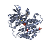
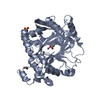
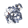
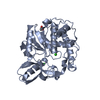
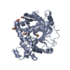
- PDB-2z8c: Phosphorylated insulin receptor tyrosine kinase in complex with (... -
+ Open data

- Basic information
| Entry | Database: PDB / ID: 2z8c | ||||||
|---|---|---|---|---|---|---|---|
| Title | Phosphorylated insulin receptor tyrosine kinase in complex with (4-{[5-carbamoyl-4-(3-methylanilino)pyrimidin-2-yl]amino}phenyl)acetic acid | ||||||
 Components Components |
| ||||||
 Keywords Keywords | TRANSFERASE / Alternative splicing / ATP-binding / Carbohydrate metabolism / Cleavage on pair of basic residues / Diabetes mellitus / Disease mutation / Glycoprotein / Kinase / Membrane / Nucleotide-binding / Phosphorylation / Polymorphism / Receptor / Transmembrane / Tyrosine-protein kinase / Transducer | ||||||
| Function / homology |  Function and homology information Function and homology informationIRS-related events triggered by IGF1R / positive regulation of fatty acid beta-oxidation / positive regulation of glucose metabolic process / regulation of female gonad development / positive regulation of meiotic cell cycle / IRS-mediated signalling / insulin-like growth factor II binding / positive regulation of developmental growth / insulin receptor complex / insulin-like growth factor I binding ...IRS-related events triggered by IGF1R / positive regulation of fatty acid beta-oxidation / positive regulation of glucose metabolic process / regulation of female gonad development / positive regulation of meiotic cell cycle / IRS-mediated signalling / insulin-like growth factor II binding / positive regulation of developmental growth / insulin receptor complex / insulin-like growth factor I binding / transmembrane receptor protein tyrosine kinase adaptor activity / Activated NTRK3 signals through PI3K / insulin receptor activity / positive regulation of protein-containing complex disassembly / Signaling by Leptin / cellular response to fatty acid / adrenal gland development / Signaling by LTK / dendritic spine maintenance / PI3K/AKT activation / insulin binding / cargo receptor activity / Signaling by ALK / PTB domain binding / Signaling by Insulin receptor / IRS activation / neuronal cell body membrane / positive regulation of respiratory burst / amyloid-beta clearance / intracellular membrane-bounded organelle / insulin receptor substrate binding / positive regulation of receptor internalization / PI3K Cascade / heart morphogenesis / regulation of embryonic development / negative regulation of insulin secretion / positive regulation of glycogen biosynthetic process / positive regulation of insulin receptor signaling pathway / Signal attenuation / SOS-mediated signalling / protein kinase activator activity / Growth hormone receptor signaling / phosphatidylinositol 3-kinase binding / transport across blood-brain barrier / Insulin receptor recycling / insulin-like growth factor receptor binding / phosphotyrosine residue binding / insulin-like growth factor receptor signaling pathway / neuron projection maintenance / negative regulation of insulin receptor signaling pathway / positive regulation of mitotic nuclear division / signaling adaptor activity / receptor-mediated endocytosis / positive regulation of glycolytic process / Insulin receptor signalling cascade / Interleukin-7 signaling / learning / SH2 domain binding / dendrite membrane / positive regulation of D-glucose import across plasma membrane / protein kinase C binding / insulin receptor binding / phosphatidylinositol 3-kinase/protein kinase B signal transduction / male gonad development / response to insulin / receptor protein-tyrosine kinase / memory / caveola / cellular response to insulin stimulus / cytokine-mediated signaling pathway / positive regulation of nitric oxide biosynthetic process / Constitutive Signaling by Aberrant PI3K in Cancer / glucose homeostasis / insulin receptor signaling pathway / Signaling by ALK fusions and activated point mutants / late endosome / PIP3 activates AKT signaling / protein autophosphorylation / amyloid-beta binding / PI5P, PP2A and IER3 Regulate PI3K/AKT Signaling / signaling receptor complex adaptor activity / RAF/MAP kinase cascade / protein tyrosine kinase activity / positive regulation of MAPK cascade / positive regulation of phosphatidylinositol 3-kinase/protein kinase B signal transduction / positive regulation of canonical NF-kappaB signal transduction / signaling receptor complex / lysosome / endosome membrane / positive regulation of cell migration / G protein-coupled receptor signaling pathway / external side of plasma membrane / protein domain specific binding / axon / positive regulation of cell population proliferation / symbiont entry into host cell / regulation of DNA-templated transcription / positive regulation of DNA-templated transcription / GTP binding / protein-containing complex binding Similarity search - Function | ||||||
| Biological species |  Homo sapiens (human) Homo sapiens (human) | ||||||
| Method |  X-RAY DIFFRACTION / SYNCHROTRON / MOLECULAR REPLACEMENT / Resolution: 3.25 Å X-RAY DIFFRACTION / SYNCHROTRON / MOLECULAR REPLACEMENT / Resolution: 3.25 Å | ||||||
 Authors Authors | Katayama, N. / Kurihara, H. | ||||||
 Citation Citation | Journal: Proteins / Year: 2008 Title: Identification of a key element for hydrogen-bonding patterns between protein kinases and their inhibitors. Authors: Katayama, N. / Orita, M. / Yamaguchi, T. / Hisamichi, H. / Kuromitsu, S. / Kurihara, H. / Sakashita, H. / Matsumoto, Y. / Fujita, S. / Niimi, T. | ||||||
| History |
|
- Structure visualization
| Structure viewer | Molecule: MolmilJmol/JSmol |
|---|
- Downloads & links
-Download
| PDBx/mmCIF format | 2z8c.cif.gz | 75.3 KB | Display | PDBx/mmCIF format |
|---|---|---|---|---|
| PDB format | pdb2z8c.ent.gz | 56 KB | Display | PDB format |
| PDBx/mmJSON format | 2z8c.json.gz | Tree view | PDBx/mmJSON format | |
| Others |  Other downloads Other downloads |
-Validation report
| Arichive directory | https://data.pdbj.org/pub/pdb/validation_reports/z8/2z8cftp://data.pdbj.org/pub/pdb/validation_reports/z8/2z8c | HTTPS FTP |
|---|
-Related structure data
| Related structure data |  2z7lC 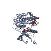 1ir3S C: citing same article ( S: Starting model for refinement |
|---|---|
| Similar structure data |


-Links
 PDBj
PDBj


- Assembly
| Deposited unit | 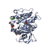
| ||||||||
|---|---|---|---|---|---|---|---|---|---|
| 1 |
| ||||||||
| Unit cell |
|
-Components
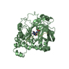
| #1: Protein | Mass: 34689.320 Da / Num. of mol.: 1 / Fragment: TYROSINE KINASE DOMAIN / Mutation: C981S, Y984F Source method: isolated from a genetically manipulated source Source: (gene. exp.) Homo sapiens (human) / Gene: INSR / Cell line (production host): SF9 / Cellular location (production host): CYTOPLASM / Production host:   SPODOPTERA FRUGIPERDA (fall armyworm) SPODOPTERA FRUGIPERDA (fall armyworm)References: UniProt: P06213, receptor protein-tyrosine kinase |
|---|---|
| #2: Protein/peptide | Mass: 759.849 Da / Num. of mol.: 1 Source method: isolated from a genetically manipulated source Source: (gene. exp.) Homo sapiens (human) / Gene: IRS1 / Cell line (production host): SF9 / Cellular location (production host): CYTOPLASM / Production host: SPODOPTERA FRUGIPERDA (fall armyworm) / References: UniProt: P35568 |
| #3: Chemical | ChemComp-S91 / [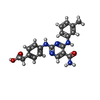  Mass: 377.397 Da / Num. of mol.: 1 / Source method: obtained synthetically / Formula: C20H19N5O3 Mass: 377.397 Da / Num. of mol.: 1 / Source method: obtained synthetically / Formula: C20H19N5O3 |
| Has protein modification | Y |
-Experimental details
-Experiment
| Experiment | Method: X-RAY DIFFRACTION / Number of used crystals: 1 |
|---|
- Sample preparation
| Crystal | Density Matthews: 2.79 Å3/Da / Density % sol: 55.88 % |
|---|---|
| Crystal grow | Method: vapor diffusion, hanging drop / Details: PEG 3350, VAPOR DIFFUSION, HANGING DROP |
-Data collection
| Diffraction source | Source: SYNCHROTRON / Site: Photon Factory  / Beamline: BL-6B / Wavelength: 1 / Beamline: BL-6B / Wavelength: 1 |
|---|---|
| Detector | Type: WEISSENBERG / Detector: DIFFRACTOMETER / Date: Jan 1, 2001 |
| Radiation | Protocol: SINGLE WAVELENGTH / Monochromatic (M) / Laue (L): M / Scattering type: x-ray |
| Radiation wavelength | Wavelength: 1 Å / Relative weight: 1 |
| Reflection | Resolution: 3.25→56.23 Å / Num. obs: 6395 / % possible obs: 98.7 % / Rmerge(I) obs: 0.092 |
- Processing
| Software |
| ||||||||||||||||||||||||||||||||||||||||||||||||||||||||||||||||||||||||||||||||
|---|---|---|---|---|---|---|---|---|---|---|---|---|---|---|---|---|---|---|---|---|---|---|---|---|---|---|---|---|---|---|---|---|---|---|---|---|---|---|---|---|---|---|---|---|---|---|---|---|---|---|---|---|---|---|---|---|---|---|---|---|---|---|---|---|---|---|---|---|---|---|---|---|---|---|---|---|---|---|---|---|---|
| Refinement | Method to determine structure: MOLECULAR REPLACEMENT Starting model: 1IR3 Resolution: 3.25→56.23 Å / Rfactor Rfree error: 0.011 / Data cutoff high absF: 1313049.57 / Data cutoff low absF: 0 / Isotropic thermal model: RESTRAINED / Cross valid method: THROUGHOUT / σ(F): 0 / Stereochemistry target values: Engh & Huber
| ||||||||||||||||||||||||||||||||||||||||||||||||||||||||||||||||||||||||||||||||
| Solvent computation | Solvent model: FLAT MODEL / Bsol: 13.78 Å2 / ksol: 0.24 e/Å3 | ||||||||||||||||||||||||||||||||||||||||||||||||||||||||||||||||||||||||||||||||
| Displacement parameters | Biso mean: 60.2 Å2
| ||||||||||||||||||||||||||||||||||||||||||||||||||||||||||||||||||||||||||||||||
| Refine analyze |
| ||||||||||||||||||||||||||||||||||||||||||||||||||||||||||||||||||||||||||||||||
| Refinement step | Cycle: LAST / Resolution: 3.25→56.23 Å
| ||||||||||||||||||||||||||||||||||||||||||||||||||||||||||||||||||||||||||||||||
| Refine LS restraints |
| ||||||||||||||||||||||||||||||||||||||||||||||||||||||||||||||||||||||||||||||||
| LS refinement shell | Resolution: 3.25→3.45 Å / Rfactor Rfree error: 0.036 / Total num. of bins used: 6
|
