 Movie
Movie Controller

[English] 日本語
 Yorodumi
- PDB-2z6s: Crystal structure of the oxy myoglobin free from X-ray-induced ph... -
+ Open data

- Basic information
| Entry | Database: PDB / ID: 2z6s | ||||||
|---|---|---|---|---|---|---|---|
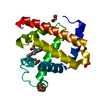
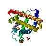
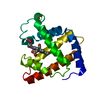
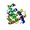
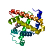
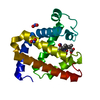
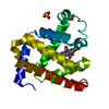
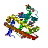
| Title | Crystal structure of the oxy myoglobin free from X-ray-induced photoreduction | ||||||
 Components Components | Myoglobin | ||||||
 Keywords Keywords | OXYGEN BINDING / heme / oxygen transport / Microspectrophotometer / X-ray-induced-photoreduction / Iron / Metal-binding / Muscle protein | ||||||
| Function / homology |  Function and homology information Function and homology informationOxidoreductases; Acting on other nitrogenous compounds as donors / nitrite reductase activity / sarcoplasm / Oxidoreductases; Acting on a peroxide as acceptor; Peroxidases / removal of superoxide radicals / oxygen carrier activity / peroxidase activity / oxygen binding / heme binding / extracellular exosome / metal ion binding Similarity search - Function | ||||||
| Biological species |  | ||||||
| Method |  X-RAY DIFFRACTION / SYNCHROTRON / MOLECULAR REPLACEMENT / Resolution: 1.25 Å X-RAY DIFFRACTION / SYNCHROTRON / MOLECULAR REPLACEMENT / Resolution: 1.25 Å | ||||||
 Authors Authors | Unno, M. / Kusama, S. / Chen, H. / Shaik, S. / Ikeda-Saito, M. | ||||||
 Citation Citation | Journal: J.Am.Chem.Soc. / Year: 2007 Title: Structural Characterization of the Fleeting Ferric Peroxo Species in Myoglobin: Experiment and Theory Authors: Unno, M. / Chen, H. / Kusama, S. / Shaik, S. / Ikeda-Saito, M. | ||||||
| History |
|
- Structure visualization
| Structure viewer | Molecule: MolmilJmol/JSmol |
|---|
- Downloads & links
-Download
| PDBx/mmCIF format | 2z6s.cif.gz | 87.9 KB | Display | PDBx/mmCIF format |
|---|---|---|---|---|
| PDB format | pdb2z6s.ent.gz | 65.7 KB | Display | PDB format |
| PDBx/mmJSON format | 2z6s.json.gz | Tree view | PDBx/mmJSON format | |
| Others |  Other downloads Other downloads |
-Validation report
| Arichive directory | https://data.pdbj.org/pub/pdb/validation_reports/z6/2z6sftp://data.pdbj.org/pub/pdb/validation_reports/z6/2z6s | HTTPS FTP |
|---|
-Related structure data
| Related structure data | 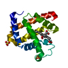 2z6tC  1am6S S: Starting model for refinement C: citing same article ( |
|---|---|
| Similar structure data |


-Links
 PDBj
PDBj


- Assembly
| Deposited unit | 
| ||||||||
|---|---|---|---|---|---|---|---|---|---|
| 1 |
| ||||||||
| Unit cell |
|
-Components
| #1: Protein | Mass: 17234.951 Da / Num. of mol.: 1 / Source method: isolated from a natural source / Source: (natural) | ||||||
|---|---|---|---|---|---|---|---|
| #2: Chemical |   Mass: 96.063 Da / Num. of mol.: 2 / Source method: obtained synthetically / Formula: SO4 Mass: 96.063 Da / Num. of mol.: 2 / Source method: obtained synthetically / Formula: SO4#3: Chemical | ChemComp-HEM / |   Mass: 616.487 Da / Num. of mol.: 1 / Source method: obtained synthetically / Formula: C34H32FeN4O4 Mass: 616.487 Da / Num. of mol.: 1 / Source method: obtained synthetically / Formula: C34H32FeN4O4#4: Chemical | ChemComp-OXY / |   Mass: 31.999 Da / Num. of mol.: 1 / Source method: obtained synthetically / Formula: O2 Mass: 31.999 Da / Num. of mol.: 1 / Source method: obtained synthetically / Formula: O2#5: Water | ChemComp-HOH / |  Mass: 18.015 Da / Num. of mol.: 177 / Source method: isolated from a natural source / Formula: H2O Mass: 18.015 Da / Num. of mol.: 177 / Source method: isolated from a natural source / Formula: H2O |
-Experimental details
-Experiment
| Experiment | Method: X-RAY DIFFRACTION / Number of used crystals: 1 |
|---|
- Sample preparation
| Crystal | Density Matthews: 1.88 Å3/Da / Density % sol: 34.46 % |
|---|---|
| Crystal grow | Temperature: 293 K / Method: liquid diffusion / pH: 6 Details: ammonium sulfate, pH6.0, LIQUID DIFFUSION, temperature 293K |
-Data collection
| Diffraction | Mean temperature: 100 K |
|---|---|
| Diffraction source | Source: SYNCHROTRON / Site: SPring-8  / Beamline: BL44B2 / Wavelength: 0.6 Å / Beamline: BL44B2 / Wavelength: 0.6 Å |
| Detector | Type: ADSC QUANTUM 210 / Detector: CCD / Date: Dec 15, 2006 |
| Radiation | Protocol: SINGLE WAVELENGTH / Monochromatic (M) / Laue (L): M / Scattering type: x-ray |
| Radiation wavelength | Wavelength: 0.6 Å / Relative weight: 1 |
| Reflection | Resolution: 1.25→50 Å / Num. obs: 35414 / % possible obs: 99.2 % / Observed criterion σ(F): 0 / Observed criterion σ(I): 0 / Redundancy: 3.6 % / Rsym value: 0.048 / Net I/σ(I): 24.4 |
| Reflection shell | Resolution: 1.25→1.29 Å / Redundancy: 3.6 % / Mean I/σ(I) obs: 2.53 / Rsym value: 0.44 / % possible all: 96.6 |
- Processing
| Software |
| |||||||||||||||||||||||||
|---|---|---|---|---|---|---|---|---|---|---|---|---|---|---|---|---|---|---|---|---|---|---|---|---|---|---|
| Refinement | Method to determine structure: MOLECULAR REPLACEMENT Starting model: 1AM6 Resolution: 1.25→8 Å / Cross valid method: Conjugated gradient / σ(F): 0 / σ(I): 0 / Stereochemistry target values: Shelx
| |||||||||||||||||||||||||
| Refinement step | Cycle: LAST / Resolution: 1.25→8 Å
|
