 Movie
Movie Controller

[English] 日本語
 Yorodumi
- PDB-2z6m: Crystal structure of Human Ferritin H8 as biotemplate for noble m... -
+ Open data

- Basic information
| Entry | Database: PDB / ID: 2z6m | ||||||
|---|---|---|---|---|---|---|---|
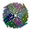
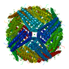
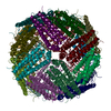
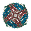
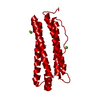
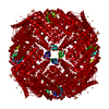
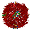
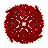
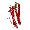
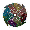
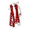
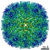
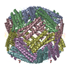
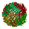
| Title | Crystal structure of Human Ferritin H8 as biotemplate for noble metal nanoparticle synthesis | ||||||
 Components Components | Ferritin heavy chain | ||||||
 Keywords Keywords | OXIDOREDUCTASE / Biotemplate / Iron / Iron storage / Metal-binding / Phosphorylation | ||||||
| Function / homology |  Function and homology information Function and homology informationiron ion sequestering activity / ferritin complex / Scavenging by Class A Receptors / Golgi Associated Vesicle Biogenesis / ferroxidase / negative regulation of ferroptosis / autolysosome / ferroxidase activity / negative regulation of fibroblast proliferation / ferric iron binding ...iron ion sequestering activity / ferritin complex / Scavenging by Class A Receptors / Golgi Associated Vesicle Biogenesis / ferroxidase / negative regulation of ferroptosis / autolysosome / ferroxidase activity / negative regulation of fibroblast proliferation / ferric iron binding / autophagosome / iron ion transport / ferrous iron binding / Iron uptake and transport / tertiary granule lumen / ficolin-1-rich granule lumen / intracellular iron ion homeostasis / immune response / iron ion binding / negative regulation of cell population proliferation / Neutrophil degranulation / extracellular exosome / extracellular region / identical protein binding / nucleus / cytoplasm / cytosol Similarity search - Function | ||||||
| Biological species |  Homo sapiens (human) Homo sapiens (human) | ||||||
| Method |  X-RAY DIFFRACTION / SYNCHROTRON / MOLECULAR REPLACEMENT / Resolution: 2.72 Å X-RAY DIFFRACTION / SYNCHROTRON / MOLECULAR REPLACEMENT / Resolution: 2.72 Å | ||||||
 Authors Authors | Di Costanzo, L. / Christianson, D.W. | ||||||
 Citation Citation | Journal: Biochemistry / Year: 2008 Title: Directing noble metal ion chemistry within a designed ferritin protein Authors: Butts, C.A. / Swift, J. / Kang, S.G. / Di Costanzo, L. / Christianson, D.W. / Saven, J.G. / Dmochowski, I.J. | ||||||
| History |
|
- Structure visualization
| Structure viewer | Molecule: MolmilJmol/JSmol |
|---|
- Downloads & links
-Download
| PDBx/mmCIF format | 2z6m.cif.gz | 419.1 KB | Display | PDBx/mmCIF format |
|---|---|---|---|---|
| PDB format | pdb2z6m.ent.gz | 345.9 KB | Display | PDB format |
| PDBx/mmJSON format | 2z6m.json.gz | Tree view | PDBx/mmJSON format | |
| Others |  Other downloads Other downloads |
-Validation report
| Arichive directory | https://data.pdbj.org/pub/pdb/validation_reports/z6/2z6mftp://data.pdbj.org/pub/pdb/validation_reports/z6/2z6m | HTTPS FTP |
|---|
-Related structure data
| Related structure data |  3erzC  3es3C  2fhaS S: Starting model for refinement C: citing same article ( |
|---|---|
| Similar structure data |


-Links
 PDBj
PDBj


- Assembly
| Deposited unit | 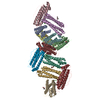
| ||||||||||||||||||
|---|---|---|---|---|---|---|---|---|---|---|---|---|---|---|---|---|---|---|---|
| 1 | 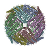
| ||||||||||||||||||
| 2 | 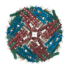
| ||||||||||||||||||
| Unit cell |
| ||||||||||||||||||
| Components on special symmetry positions |
|
-Components
| #1: Protein | Mass: 20361.869 Da / Num. of mol.: 12 / Fragment: residues 1-176 Mutation: H13D, E64C, C90R, C102A, H105Q, E140C, K143C, E147C Source method: isolated from a genetically manipulated source Source: (gene. exp.) Homo sapiens (human) / Production host:  #2: Chemical | ChemComp-ZN /   Mass: 65.409 Da / Num. of mol.: 5 / Source method: obtained synthetically / Formula: Zn Mass: 65.409 Da / Num. of mol.: 5 / Source method: obtained synthetically / Formula: Zn#3: Chemical | ChemComp-CA /   Mass: 40.078 Da / Num. of mol.: 4 / Source method: obtained synthetically / Formula: Ca Mass: 40.078 Da / Num. of mol.: 4 / Source method: obtained synthetically / Formula: Ca#4: Chemical | ChemComp-MPD / (   Mass: 118.174 Da / Num. of mol.: 7 / Source method: obtained synthetically / Formula: C6H14O2 / Comment: precipitant*YM Mass: 118.174 Da / Num. of mol.: 7 / Source method: obtained synthetically / Formula: C6H14O2 / Comment: precipitant*YM#5: Water | ChemComp-HOH / |  Mass: 18.015 Da / Num. of mol.: 368 / Source method: isolated from a natural source / Formula: H2O Mass: 18.015 Da / Num. of mol.: 368 / Source method: isolated from a natural source / Formula: H2O |
|---|
-Experimental details
-Experiment
| Experiment | Method: X-RAY DIFFRACTION / Number of used crystals: 1 |
|---|
- Sample preparation
| Crystal | Density Matthews: 2.86 Å3/Da / Density % sol: 56.98 % |
|---|---|
| Crystal grow | Temperature: 298 K / Method: vapor diffusion, hanging drop / pH: 4.6 Details: protein solution(11.0 mg/mL H8 in unbuffered 3.0mM NaN3), 2.5mL of precipitant buffer(0.1 M sodium acetate(pH 4.6), 20%(v/v) isopropanol, 0.2M CaCl2) , VAPOR DIFFUSION, HANGING DROP, temperature 298K |
-Data collection
| Diffraction | Mean temperature: 100 K |
|---|---|
| Diffraction source | Source: SYNCHROTRON / Site: NSLS  / Beamline: X29A / Wavelength: 1.08 Å / Beamline: X29A / Wavelength: 1.08 Å |
| Detector | Type: ADSC QUANTUM 315 / Detector: CCD / Date: Jul 28, 2007 |
| Radiation | Protocol: SINGLE WAVELENGTH / Monochromatic (M) / Laue (L): M / Scattering type: x-ray |
| Radiation wavelength | Wavelength: 1.08 Å / Relative weight: 1 |
| Reflection | Resolution: 2.72→50 Å / Num. obs: 75646 / % possible obs: 99 % / Observed criterion σ(I): 2 / Redundancy: 3.6 % / Rmerge(I) obs: 0.096 / Net I/σ(I): 8.8 |
| Reflection shell | Resolution: 2.72→2.82 Å / Redundancy: 3.4 % / Rmerge(I) obs: 0.4 / Mean I/σ(I) obs: 1.9 / Num. unique all: 7408 / % possible all: 98.7 |
- Processing
| Software |
| ||||||||||||||||||||
|---|---|---|---|---|---|---|---|---|---|---|---|---|---|---|---|---|---|---|---|---|---|
| Refinement | Method to determine structure: MOLECULAR REPLACEMENT Starting model: 2FHA Resolution: 2.72→50 Å / σ(F): 0 / σ(I): 1
| ||||||||||||||||||||
| Refinement step | Cycle: LAST / Resolution: 2.72→50 Å
| ||||||||||||||||||||
| Refine LS restraints |
| ||||||||||||||||||||
| LS refinement shell | Resolution: 2.72→2.82 Å / Rfactor Rfree: 0.357 / Rfactor Rwork: 0.317 |
