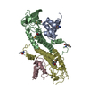
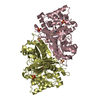
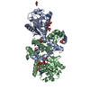
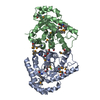
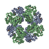
Crystal Structure of 5-aminolevulinic acid dehydratase (ALAD) from Mus musculus
Components
Delta-aminolevulinic acid dehydratase
Keywords
LYASE / DEHYDRATASE / TETRAPYRROLE BIOSYNTHESIS / Structural Genomics / NPPSFA / National Project on Protein Structural and Functional Analyses / RIKEN Structural Genomics/Proteomics Initiative / RSGI
Function / homology
Function and homology information
Heme biosynthesis / proteasome core complex binding / response to vitamin B1 / cellular response to lead ion / response to platinum ion / porphobilinogen synthase / porphobilinogen synthase activity / heme A biosynthetic process / response to aluminum ion / negative regulation of proteasomal protein catabolic process ...Heme biosynthesis / proteasome core complex binding / response to vitamin B1 / cellular response to lead ion / response to platinum ion / porphobilinogen synthase / porphobilinogen synthase activity / heme A biosynthetic process / response to aluminum ion / negative regulation of proteasomal protein catabolic process / heme B biosynthetic process / response to mercury ion / : / response to selenium ion / response to cobalt ion / response to fatty acid / response to methylmercury / response to arsenic-containing substance / response to iron ion / heme biosynthetic process / response to herbicide / response to metal ion / response to zinc ion / response to vitamin E / response to ionizing radiation / response to amino acid / response to cadmium ion / Neutrophil degranulation / response to nutrient / cellular response to interleukin-4 / response to glucocorticoid / response to activity / protein homooligomerization / response to oxidative stress / response to lipopolysaccharide / response to ethanol / response to hypoxia / response to xenobiotic stimulus / : / zinc ion binding / metal ion binding / identical protein binding / cytosol Similarity search - Function
Delta-aminolevulinic acid dehydratase / Delta-aminolevulinic acid dehydratase, active site / Delta-aminolevulinic acid dehydratase / Delta-aminolevulinic acid dehydratase active site. / Delta-aminolevulinic acid dehydratase / Aldolase class I / Aldolase-type TIM barrel / TIM Barrel / Alpha-Beta Barrel / Alpha Beta Similarity search - Domain/homology
In the structure databanks used in Yorodumi, some data are registered as the other names, "COVID-19 virus" and "2019-nCoV". Here are the details of the virus and the list of structure data.
Jan 31, 2019. EMDB accession codes are about to change! (news from PDBe EMDB page)
EMDB accession codes are about to change! (news from PDBe EMDB page)
The allocation of 4 digits for EMDB accession codes will soon come to an end. Whilst these codes will remain in use, new EMDB accession codes will include an additional digit and will expand incrementally as the available range of codes is exhausted. The current 4-digit format prefixed with “EMD-” (i.e. EMD-XXXX) will advance to a 5-digit format (i.e. EMD-XXXXX), and so on. It is currently estimated that the 4-digit codes will be depleted around Spring 2019, at which point the 5-digit format will come into force.
The EM Navigator/Yorodumi systems omit the EMD- prefix.
Related info.:Q: What is EMD? / ID/Accession-code notation in Yorodumi/EM Navigator
Yorodumi is a browser for structure data from EMDB, PDB, SASBDB, etc.
This page is also the successor to EM Navigator detail page, and also detail information page/front-end page for Omokage search.
The word "yorodu" (or yorozu) is an old Japanese word meaning "ten thousand". "mi" (miru) is to see.
Related info.:EMDB / PDB / SASBDB / Comparison of 3 databanks / Yorodumi Search / Aug 31, 2016. New EM Navigator & Yorodumi / Yorodumi Papers / Jmol/JSmol / Function and homology information / Changes in new EM Navigator and Yorodumi
 Movie
Movie Controller

 Yorodumi
 Open data
 Basic information
 Components
 Keywords
 Function and homology information
 X-RAY DIFFRACTION /
  Authors
 Citation
 Structure visualization
 Downloads & links
 Other downloads


 PDBj
PDBj


 Assembly
 Mass: 18.015 Da / Num. of mol.: 48 / Source method: isolated from a natural source / Formula: H2O
 Sample preparation
 / Beamline: BL-5A / Wavelength: 0.97931 Å
 Processing