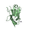
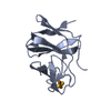
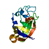
METAL BINDING PROTEIN / zf-B_box domain / Structural Genomics / NPPSFA / National Project on Protein Structural and Functional Analyses / RIKEN Structural Genomics/Proteomics Initiative / RSGI
Function / homology
Function and homology information
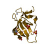
convergent extension involved in axis elongation / Krueppel-associated box domain binding / embryonic placenta morphogenesis / negative regulation of single stranded viral RNA replication via double stranded DNA intermediate / chromo shadow domain binding / suppression of viral release by host / genomic imprinting / Generic Transcription Pathway / SUMO transferase activity / DNA methylation-dependent constitutive heterochromatin formation ...convergent extension involved in axis elongation / Krueppel-associated box domain binding / embryonic placenta morphogenesis / negative regulation of single stranded viral RNA replication via double stranded DNA intermediate / chromo shadow domain binding / suppression of viral release by host / genomic imprinting / Generic Transcription Pathway / SUMO transferase activity / DNA methylation-dependent constitutive heterochromatin formation / epithelial to mesenchymal transition / protein sumoylation / heterochromatin / SUMOylation of transcription cofactors / embryo implantation / positive regulation of DNA repair / Regulation of endogenous retroelements by KRAB-ZFP proteins / promoter-specific chromatin binding / euchromatin / positive regulation of protein import into nucleus / RING-type E3 ubiquitin transferase / RNA polymerase II transcription regulator complex / HCMV Early Events / ubiquitin-protein transferase activity / transcription corepressor activity / ubiquitin protein ligase activity / chromatin organization / proteasome-mediated ubiquitin-dependent protein catabolic process / protein kinase activity / transcription coactivator activity / innate immune response / DNA repair / negative regulation of DNA-templated transcription / chromatin binding / ubiquitin protein ligase binding / positive regulation of DNA-templated transcription / negative regulation of transcription by RNA polymerase II / protein-containing complex / DNA binding / RNA binding / zinc ion binding / nucleoplasm / nucleus Similarity search - Function
In the structure databanks used in Yorodumi, some data are registered as the other names, "COVID-19 virus" and "2019-nCoV". Here are the details of the virus and the list of structure data.
Jan 31, 2019. EMDB accession codes are about to change! (news from PDBe EMDB page)
EMDB accession codes are about to change! (news from PDBe EMDB page)
The allocation of 4 digits for EMDB accession codes will soon come to an end. Whilst these codes will remain in use, new EMDB accession codes will include an additional digit and will expand incrementally as the available range of codes is exhausted. The current 4-digit format prefixed with “EMD-” (i.e. EMD-XXXX) will advance to a 5-digit format (i.e. EMD-XXXXX), and so on. It is currently estimated that the 4-digit codes will be depleted around Spring 2019, at which point the 5-digit format will come into force.
The EM Navigator/Yorodumi systems omit the EMD- prefix.
Related info.:Q: What is EMD? / ID/Accession-code notation in Yorodumi/EM Navigator
Yorodumi is a browser for structure data from EMDB, PDB, SASBDB, etc.
This page is also the successor to EM Navigator detail page, and also detail information page/front-end page for Omokage search.
The word "yorodu" (or yorozu) is an old Japanese word meaning "ten thousand". "mi" (miru) is to see.
Related info.:EMDB / PDB / SASBDB / Comparison of 3 databanks / Yorodumi Search / Aug 31, 2016. New EM Navigator & Yorodumi / Yorodumi Papers / Jmol/JSmol / Function and homology information / Changes in new EM Navigator and Yorodumi
 Movie
Movie Controller


 Open data
 Basic information
 Components
 Keywords
 Function and homology information
 Homo sapiens (human)
 X-RAY DIFFRACTION /
  Authors
 Citation
 Structure visualization
 Downloads & links
 Other downloads


 PDBj
PDBj

 Assembly

 Mass: 65.409 Da / Num. of mol.: 4 / Source method: obtained synthetically / Formula: Zn
 Mass: 18.015 Da / Num. of mol.: 95 / Source method: isolated from a natural source / Formula: H2O
 Sample preparation
 / Beamline: BL26B2 / Wavelength: 1.2820, 1.2831
 Processing