[English] 日本語
 Yorodumi
- PDB-2ynu: Apo GIM-1 with 2Mol. Crystal structures of Pseudomonas aeruginosa... -
+ Open data
- Basic information
| Entry | Database: PDB / ID: 2ynu | ||||||
|---|---|---|---|---|---|---|---|
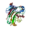
| Title | Apo GIM-1 with 2Mol. Crystal structures of Pseudomonas aeruginosa GIM-1: active site plasticity in metallo-beta-lactamases | ||||||
 Components Components | GIM-1 PROTEIN | ||||||
 Keywords Keywords | HYDROLASE / ANTIBIOTIC RESISTANCE / RESIDUE DETERMINANTS / LOOP DYNAMICS | ||||||
| Function / homology |  Function and homology information Function and homology information | ||||||
| Biological species |  | ||||||
| Method |  X-RAY DIFFRACTION / X-RAY DIFFRACTION /  SYNCHROTRON / SYNCHROTRON /  MOLECULAR REPLACEMENT / Resolution: 2.06 Å MOLECULAR REPLACEMENT / Resolution: 2.06 Å | ||||||
 Authors Authors | Borra, P.S. / Samuelsen, O. / Spencer, J. / Lorentzen, M.S. / Leiros, H.-K.S. | ||||||
 Citation Citation |  Journal: Antimicrob.Agents Chemother. / Year: 2013 Journal: Antimicrob.Agents Chemother. / Year: 2013Title: Crystal Structures of Pseudomonas Aeruginosa Gim-1: Active-Site Plasticity in Metallo-Beta-Lactamases. Authors: Borra, P.S. / Samuelsen, O. / Spencer, J. / Walsh, T.R. / Lorentzen, M.S. / Leiros, H.-K.S. | ||||||
| History |
|
- Structure visualization
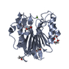
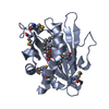
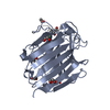
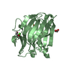
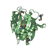
| Structure viewer | Molecule:  Molmil Molmil Jmol/JSmol Jmol/JSmol |
|---|
- Downloads & links
- Download
| PDBx/mmCIF format |  2ynu.cif.gz 2ynu.cif.gz | 171.9 KB | Display |  PDBx/mmCIF format PDBx/mmCIF format |
|---|---|---|---|---|
| PDB format |  pdb2ynu.ent.gz pdb2ynu.ent.gz | 138.9 KB | Display |  PDB format PDB format |
| PDBx/mmJSON format |  2ynu.json.gz 2ynu.json.gz | Tree view |  PDBx/mmJSON format PDBx/mmJSON format | |
| Others |  Other downloads Other downloads |
-Validation report
| Summary document |  2ynu_validation.pdf.gz 2ynu_validation.pdf.gz | 435.5 KB | Display |  wwPDB validaton report wwPDB validaton report |
|---|---|---|---|---|
| Full document |  2ynu_full_validation.pdf.gz 2ynu_full_validation.pdf.gz | 446 KB | Display | |
| Data in XML |  2ynu_validation.xml.gz 2ynu_validation.xml.gz | 20.3 KB | Display | |
| Data in CIF |  2ynu_validation.cif.gz 2ynu_validation.cif.gz | 28.3 KB | Display | |
| Arichive directory |  https://data.pdbj.org/pub/pdb/validation_reports/yn/2ynu https://data.pdbj.org/pub/pdb/validation_reports/yn/2ynu ftp://data.pdbj.org/pub/pdb/validation_reports/yn/2ynu ftp://data.pdbj.org/pub/pdb/validation_reports/yn/2ynu | HTTPS FTP |
-Related structure data
- Links
Links
- Assembly
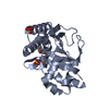
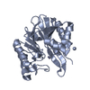
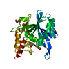
| Deposited unit | 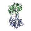
| ||||||||
|---|---|---|---|---|---|---|---|---|---|
| 1 | 
| ||||||||
| 2 | 
| ||||||||
| Unit cell |
|
- Components
| #1: Protein | Mass: 25619.758 Da / Num. of mol.: 2 Source method: isolated from a genetically manipulated source Source: (gene. exp.)   #2: Water | ChemComp-HOH / | Sequence details | RESIDUES 1-18 IN THE GENE SEQUENCE ARE REMOVED AND A HIS TAG AND TEV CLEAVE SITE WAS INTRODUCED. A ...RESIDUES 1-18 IN THE GENE SEQUENCE ARE REMOVED AND A HIS TAG AND TEV CLEAVE SITE WAS INTRODUCED | |
|---|
-Experimental details
-Experiment
| Experiment | Method:  X-RAY DIFFRACTION X-RAY DIFFRACTION |
|---|
- Sample preparation
| Crystal | Density Matthews: 2.05 Å3/Da / Density % sol: 39.91 % / Description: NONE |
|---|---|
| Crystal grow | Details: 0.1M TRIS PH 7.1, 21% POLYETHYLENE GLYCOL MONOMETHYL ETHER 2000 (PEG MME 2K), 4% GLYCEROL |
-Data collection
| Diffraction | Mean temperature: 100 K |
|---|---|
| Diffraction source | Source:  SYNCHROTRON / Site: SYNCHROTRON / Site:  ESRF ESRF  / Beamline: ID29 / Wavelength: 0.97239 / Beamline: ID29 / Wavelength: 0.97239 |
| Detector | Type: ADSC CCD / Detector: CCD |
| Radiation | Protocol: SINGLE WAVELENGTH / Monochromatic (M) / Laue (L): M / Scattering type: x-ray |
| Radiation wavelength | Wavelength: 0.97239 Å / Relative weight: 1 |
| Reflection | Resolution: 2.06→45 Å / Num. obs: 25067 / % possible obs: 97.2 % / Observed criterion σ(I): 0 / Redundancy: 2.8 % / Rmerge(I) obs: 0.04 / Net I/σ(I): 12.1 |
| Reflection shell | Resolution: 2.06→2.17 Å / Redundancy: 2.8 % / Rmerge(I) obs: 0.57 / Mean I/σ(I) obs: 2 / % possible all: 98.7 |
- Processing
| Software |
| ||||||||||||||||||||||||||||||||||||||||||||||||||||||||||||||||||||||
|---|---|---|---|---|---|---|---|---|---|---|---|---|---|---|---|---|---|---|---|---|---|---|---|---|---|---|---|---|---|---|---|---|---|---|---|---|---|---|---|---|---|---|---|---|---|---|---|---|---|---|---|---|---|---|---|---|---|---|---|---|---|---|---|---|---|---|---|---|---|---|---|
| Refinement | Method to determine structure:  MOLECULAR REPLACEMENT / Resolution: 2.06→19.712 Å / SU ML: 0.3 / σ(F): 1.36 / Phase error: 28.03 / Stereochemistry target values: ML MOLECULAR REPLACEMENT / Resolution: 2.06→19.712 Å / SU ML: 0.3 / σ(F): 1.36 / Phase error: 28.03 / Stereochemistry target values: ML
| ||||||||||||||||||||||||||||||||||||||||||||||||||||||||||||||||||||||
| Solvent computation | Shrinkage radii: 0.9 Å / VDW probe radii: 1.11 Å / Solvent model: FLAT BULK SOLVENT MODEL | ||||||||||||||||||||||||||||||||||||||||||||||||||||||||||||||||||||||
| Refinement step | Cycle: LAST / Resolution: 2.06→19.712 Å
| ||||||||||||||||||||||||||||||||||||||||||||||||||||||||||||||||||||||
| Refine LS restraints |
| ||||||||||||||||||||||||||||||||||||||||||||||||||||||||||||||||||||||
| LS refinement shell |
|
 Movie
Movie Controller



 PDBj
PDBj


