 Movie
Movie Controller

[English] 日本語
 Yorodumi
- PDB-2yjt: Crystal structure of E. coli DEAD-box protein SrmB bound to regul... -
+ Open data

- Basic information
| Entry | Database: PDB / ID: 2yjt | ||||||
|---|---|---|---|---|---|---|---|
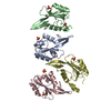
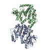
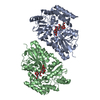
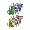
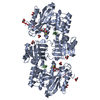
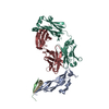
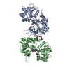
| Title | Crystal structure of E. coli DEAD-box protein SrmB bound to regulator of ribonuclease activity A (RraA) | ||||||
 Components Components |
| ||||||
 Keywords Keywords | HYDROLASE INHIBITOR/HYDROLASE / HYDROLASE INHIBITOR-HYDROLASE COMPLEX / DEAD BOX RNA HELICASES | ||||||
| Function / homology |  Function and homology information Function and homology informationnegative regulation of RNA catabolic process / ribonuclease inhibitor activity / RNA strand annealing activity / poly(A) binding / ATP-dependent activity, acting on RNA / protein homotrimerization / transcriptional attenuation / endoribonuclease inhibitor activity / ribosomal large subunit assembly / RNA helicase activity ...negative regulation of RNA catabolic process / ribonuclease inhibitor activity / RNA strand annealing activity / poly(A) binding / ATP-dependent activity, acting on RNA / protein homotrimerization / transcriptional attenuation / endoribonuclease inhibitor activity / ribosomal large subunit assembly / RNA helicase activity / RNA helicase / enzyme binding / ATP hydrolysis activity / protein-containing complex / ATP binding / identical protein binding / cytosol Similarity search - Function | ||||||
| Biological species |  | ||||||
| Method |  X-RAY DIFFRACTION / SYNCHROTRON / MOLECULAR REPLACEMENT / Resolution: 2.9 Å X-RAY DIFFRACTION / SYNCHROTRON / MOLECULAR REPLACEMENT / Resolution: 2.9 Å | ||||||
 Authors Authors | Pietras, Z. / Hardwick, S.W. / Luisi, B.F. | ||||||
 Citation Citation | Journal: J.Biol.Chem. / Year: 2013 Title: Potential Regulatory Interactions of Escherichia Coli Rraa Protein with Dead-Box Helicases. Authors: Pietras, Z. / Hardwick, S.W. / Swiezewski, S. / Luisi, B.F. | ||||||
| History |
|
- Structure visualization
| Structure viewer | Molecule: MolmilJmol/JSmol |
|---|
- Downloads & links
-Download
| PDBx/mmCIF format | 2yjt.cif.gz | 133.1 KB | Display | PDBx/mmCIF format |
|---|---|---|---|---|
| PDB format | pdb2yjt.ent.gz | 104.9 KB | Display | PDB format |
| PDBx/mmJSON format | 2yjt.json.gz | Tree view | PDBx/mmJSON format | |
| Others |  Other downloads Other downloads |
-Validation report
| Arichive directory | https://data.pdbj.org/pub/pdb/validation_reports/yj/2yjtftp://data.pdbj.org/pub/pdb/validation_reports/yj/2yjt | HTTPS FTP |
|---|
-Related structure data
| Related structure data | 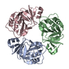 2yjvC 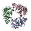 1q5xS C: citing same article ( S: Starting model for refinement |
|---|---|
| Similar structure data |

-Links
 PDBj
PDBj- Assembly
| Deposited unit | 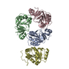
| ||||||||||||||||||||||||
|---|---|---|---|---|---|---|---|---|---|---|---|---|---|---|---|---|---|---|---|---|---|---|---|---|---|
| 1 |
| ||||||||||||||||||||||||
| Unit cell |
| ||||||||||||||||||||||||
| Noncrystallographic symmetry (NCS) | NCS domain:
NCS domain segments: Component-ID: 1 / Ens-ID: 1 / Beg auth comp-ID: LYS / Beg label comp-ID: LYS / End auth comp-ID: ASP / End label comp-ID: ASP / Refine code: 4 / Auth seq-ID: 2 - 159 / Label seq-ID: 2 - 159
|
-Components
| #1: Protein | Mass: 17371.264 Da / Num. of mol.: 3 Source method: isolated from a genetically manipulated source Source: (gene. exp.) #2: Protein | | Mass: 19559.531 Da / Num. of mol.: 1 / Fragment: RESIDUES 219-388 Source method: isolated from a genetically manipulated source Source: (gene. exp.) #3: Water | ChemComp-HOH / |  Mass: 18.015 Da / Num. of mol.: 29 / Source method: isolated from a natural source / Formula: H2O Mass: 18.015 Da / Num. of mol.: 29 / Source method: isolated from a natural source / Formula: H2O |
|---|
-Experimental details
-Experiment
| Experiment | Method: X-RAY DIFFRACTION / Number of used crystals: 1 |
|---|
- Sample preparation
| Crystal | Density Matthews: 2.47 Å3/Da / Density % sol: 50 % / Description: NONE |
|---|---|
| Crystal grow | Temperature: 289 K / Method: vapor diffusion, hanging drop / pH: 7.2 Details: CRYSTALS WERE PREPARED USING THE HANGING DROP METHOD AT 16 DEG. C BY MIXING 1:1 PROTEIN SAMPLE WITH MOTHER LIQUOR: 100 MM MAGNESIUM ACETATE, 100 MM MOPS PH 7.2 AND 12% W/V PEG 8000.THE ...Details: CRYSTALS WERE PREPARED USING THE HANGING DROP METHOD AT 16 DEG. C BY MIXING 1:1 PROTEIN SAMPLE WITH MOTHER LIQUOR: 100 MM MAGNESIUM ACETATE, 100 MM MOPS PH 7.2 AND 12% W/V PEG 8000.THE CRYSTALS WERE TRANSFERRED BRIEFLY TO RESERVOIR SOLUTION SUPPLEMENTED WITH 25% (V/V) GLYCEROL AND FLASH FROZEN IN LIQUID NITROGEN. |
-Data collection
| Diffraction | Mean temperature: 100 K |
|---|---|
| Diffraction source | Source: SYNCHROTRON / Site: Diamond  / Beamline: I04 / Wavelength: 0.9763 / Beamline: I04 / Wavelength: 0.9763 |
| Detector | Type: ADSC CCD / Detector: CCD / Date: Jan 29, 2010 / Details: MIRRORS |
| Radiation | Monochromator: SILICON CRYSTAL / Protocol: SINGLE WAVELENGTH / Monochromatic (M) / Laue (L): M / Scattering type: x-ray |
| Radiation wavelength | Wavelength: 0.9763 Å / Relative weight: 1 |
| Reflection | Resolution: 2.9→63.56 Å / Num. obs: 15208 / % possible obs: 99.6 % / Observed criterion σ(I): 6 / Redundancy: 4 % / Rmerge(I) obs: 0.12 / Net I/σ(I): 10.2 |
| Reflection shell | Resolution: 2.9→3.06 Å / Redundancy: 4.1 % / Rmerge(I) obs: 0.51 / Mean I/σ(I) obs: 2.8 / % possible all: 99.5 |
- Processing
| Software |
| ||||||||||||||||||||||||||||||||||||||||||||||||||||||||||||||||||||||||||||||||||||||||||||||||||||||||||||||||||||||||||||||||||||||||||||||||||||||||||||||||||||||||||||||||||||||
|---|---|---|---|---|---|---|---|---|---|---|---|---|---|---|---|---|---|---|---|---|---|---|---|---|---|---|---|---|---|---|---|---|---|---|---|---|---|---|---|---|---|---|---|---|---|---|---|---|---|---|---|---|---|---|---|---|---|---|---|---|---|---|---|---|---|---|---|---|---|---|---|---|---|---|---|---|---|---|---|---|---|---|---|---|---|---|---|---|---|---|---|---|---|---|---|---|---|---|---|---|---|---|---|---|---|---|---|---|---|---|---|---|---|---|---|---|---|---|---|---|---|---|---|---|---|---|---|---|---|---|---|---|---|---|---|---|---|---|---|---|---|---|---|---|---|---|---|---|---|---|---|---|---|---|---|---|---|---|---|---|---|---|---|---|---|---|---|---|---|---|---|---|---|---|---|---|---|---|---|---|---|---|---|
| Refinement | Method to determine structure: MOLECULAR REPLACEMENT Starting model: PDB ENTRY 1Q5X Resolution: 2.9→74.3 Å / Cor.coef. Fo:Fc: 0.928 / Cor.coef. Fo:Fc free: 0.882 / SU B: 16.027 / SU ML: 0.305 / Cross valid method: THROUGHOUT / ESU R Free: 0.435 / Stereochemistry target values: MAXIMUM LIKELIHOOD / Details: HYDROGENS HAVE BEEN ADDED IN THE RIDING POSITIONS.
| ||||||||||||||||||||||||||||||||||||||||||||||||||||||||||||||||||||||||||||||||||||||||||||||||||||||||||||||||||||||||||||||||||||||||||||||||||||||||||||||||||||||||||||||||||||||
| Displacement parameters | Biso mean: 39.431 Å2
| ||||||||||||||||||||||||||||||||||||||||||||||||||||||||||||||||||||||||||||||||||||||||||||||||||||||||||||||||||||||||||||||||||||||||||||||||||||||||||||||||||||||||||||||||||||||
| Refinement step | Cycle: LAST / Resolution: 2.9→74.3 Å
| ||||||||||||||||||||||||||||||||||||||||||||||||||||||||||||||||||||||||||||||||||||||||||||||||||||||||||||||||||||||||||||||||||||||||||||||||||||||||||||||||||||||||||||||||||||||
| Refine LS restraints |
|